Japanese
English
- 有料閲覧
- Abstract 文献概要
- 1ページ目 Look Inside
小脳は,発生学的に,古小脳archicerebellum,旧小脳paleocerebellum,新小脳neocerebellumの三つの部分に分けられる。これらは,それぞれ主として前庭,脊髄,大脳皮質からの投射を受ける部位に相当する。小脳皮質と小脳核との結合関係も考慮して,小脳を内側部medial part,中間部intermcdiate part,外側部lateral partに分けることもある。臨床的には,小脳虫部vermisと半球部hemisphereに分け,これに小脳脚を加えて,それぞれの部位の病変に対応して症状を考える方が現実的なことが多い。
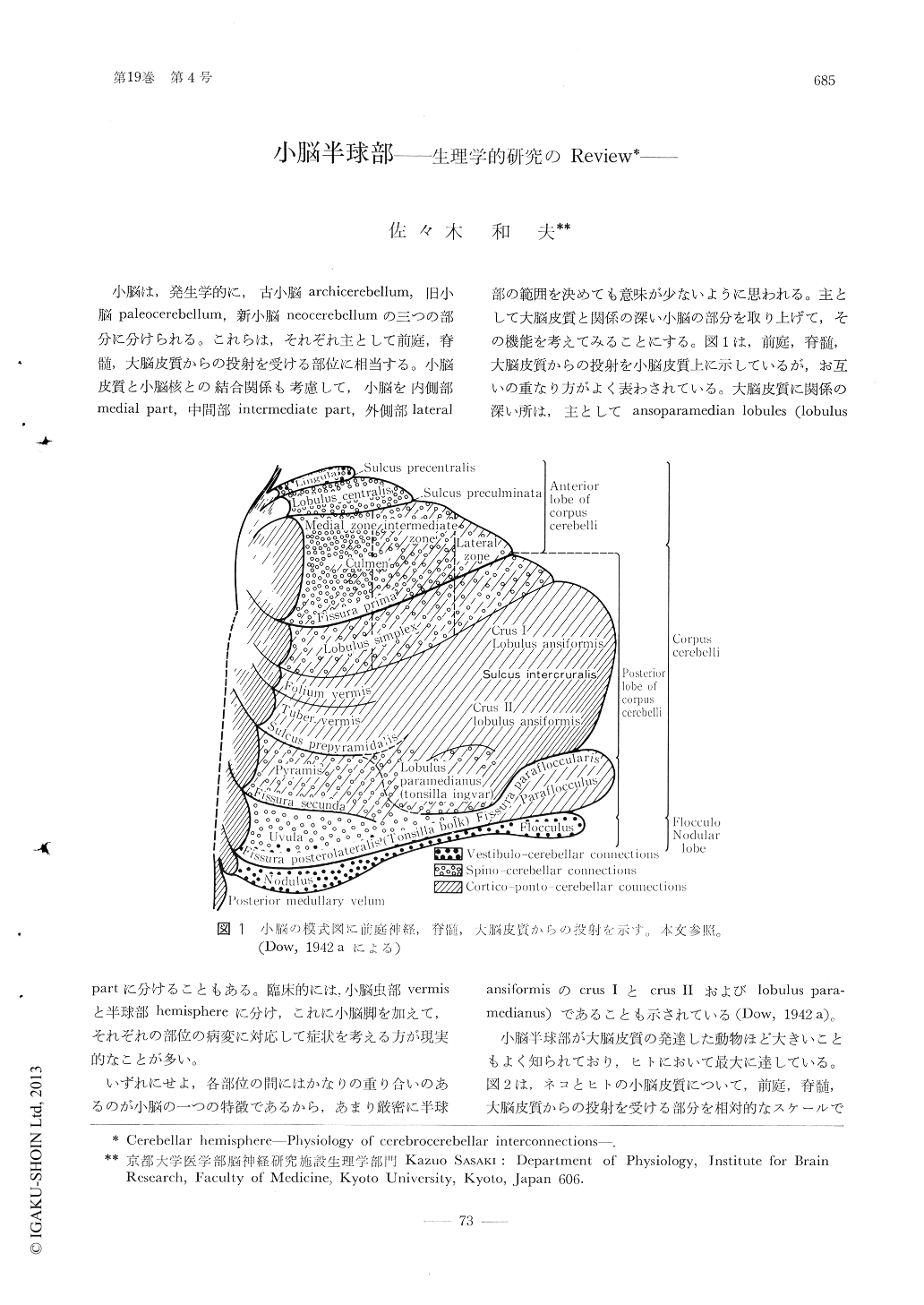
いずれにせよ,各部位の間にはかなりの重り合いのあるのが小脳の一つの特徴であるから,あまり厳密に半球部の範囲を決めても意味が少ないように思われる。主として大脳皮質と関係の深い小脳の部分を取り上げて,その機能を考えてみることにする。図1は,前庭,脊髄,大脳皮質からの投射を小脳皮質上に示しているが,お互いの重なり方がよく表わされている。大脳皮質に関係の深い所は,主としてansoparamedian lobules(lobulus ansiformisのcrus Ⅰとcrus Ⅱおよびlobulus paramedianus)であることも示されている(Dow,1942 a)。
Ablation and stimulation experiments of the cerebellar hemisphere were reviewed and summarized in referring to clinical cerebellar syndromes. Differences between the syndromes of total ablation of the cerebellum in subprimates and primates are conspicuous im muscle tone, i.e., cats and dogs show marked opisthotonos and extensor muscle rigidity for several days after the ablation, whereas monkeys, chimpanzees and humans are reported to reveal atonia and asthenia for a few days or more after lesion of the total cerebellum.

Copyright © 1975, Igaku-Shoin Ltd. All rights reserved.


