Japanese
English
- 有料閲覧
- Abstract 文献概要
- 1ページ目 Look Inside
緒言
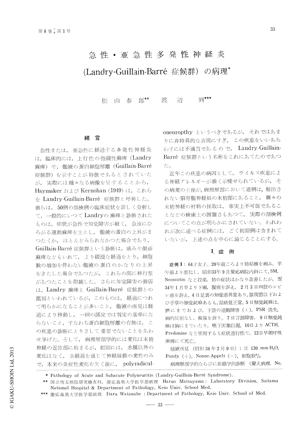
急性または,亜急性に経過する多発性神経炎は,臨床的には,上行性の弛緩性麻癖(Landry麻痺)や,髄液の蛋白細胞解離(Guillain-Barré症候群)を示すことが特徴であるとされていたが,実際には種々なる病像を呈することから,HaymakerおよびKernohan(1949)は,これらをLandry-Guillain-Barré症候群と呼称した。彼らは,50例の剖検例の臨床症状を詳しく分析して,一般的にいつてLandryの麻痺と診断されたものは,症状が急性で知覚障害が軽く,急速にひろがる運動麻痺を主とし,髄液の蛋白の上昇がまつたくか,ほとんどみられたかつた場合であり,Guillain-Barré症候群という診断は,痛みや顔面麻痺などもいれて,より緩漫な経過をとり,細胞数の増加を伴わない髄液の蛋白のかなりの上昇をきたした場合であつたが,これらの間に移行型があつたことを指摘した。さらに知覚障害の強弱は,Landry麻痺とGuillain-Barré症候群との鑑別といわれているが,このものは,経過につれて明らかになることが多いこと,髄液の所見は経過により移動し,一回の測定では判定の基灘にたらないこと,すなわち蛋白細胞解離の有無は,この疾患の診断にとりさして重要でないことをあわせ挙げた。
5 cases of acute and subacute disseminated (polyradiculo) neuritis, duration of the illness ranging from 10 days to 2 months, were studied histopathologically.
Principal change is disintegration of the myelin in the Schwann cells and the axon and ganglion cells are relatively well preserved. Together with etiopathogenesis these changes are compatible with that of the demylinative diseases in CNS. Only difference is that demylinative lesion in CNS is replaced by glia scar while in PNS the lesion is easily remyelinated completely.
There were various ages of lesions; lymphocytic infiltration around small vessels from where pleomorphic cells permeate into nervous tissus with or without demyelination, more dense accumulations of the mononuclear cells always associated with demyelination, granular disintegration of myelin in the Schwann cells without any inflammatory cell response, maked Schwann cell proliferation with mitosis and at last remyelination.
Intensity of lesions also varied and a number of minute foci of necrosis consisted of destruction of whole nervous tissue associated with mononuclear cell and macrophage response. Blood imbibition and other circulatory disturbances could be noted. Then secondary degeneration follows.
Scattered or focally accumulated karyorrhexis or nuclear debris in immediately vicinity of the Schwann cell containing myelin debris found in one case may suggest tha tthe pleomorphic wandering cells, of which early lesions seemed to be composed, have been disintegrated at the time of demyelination. Segmental demyelination itself appeared to not provoke inflammatory cell response unlesswhole ne rvous tissue has been broken down. After complete demyelination, therefore, it may be little, if any, inflammatory cells to be associated.
The lesion appears to start from the region where the nerve roots penetrate the arachnoid memrane and are enwrapped with the dura down to the region the latter transit to the periost. Especially in the spinal ganglia frequently were noted older lesions such as diffuse area of marked Schwann cell proliferation. Then lesions disseminated centrally and periferally often showing diffuse involvement extending to subperineurial or subpial region with occasional severe destruction of nervous tissue.
Edematous process was more or less associated with damage of the nervous tissue and endoneural connective tissue seemed to increase without apparent fibroblastic activity. In severe lesion other circulatory disturbance such as blood imbibition also occurred. These may serve to accentuate foci and the fuse each other.

Copyright © 1964, Igaku-Shoin Ltd. All rights reserved.


