Japanese
English
- 有料閲覧
- Abstract 文献概要
- 1ページ目 Look Inside
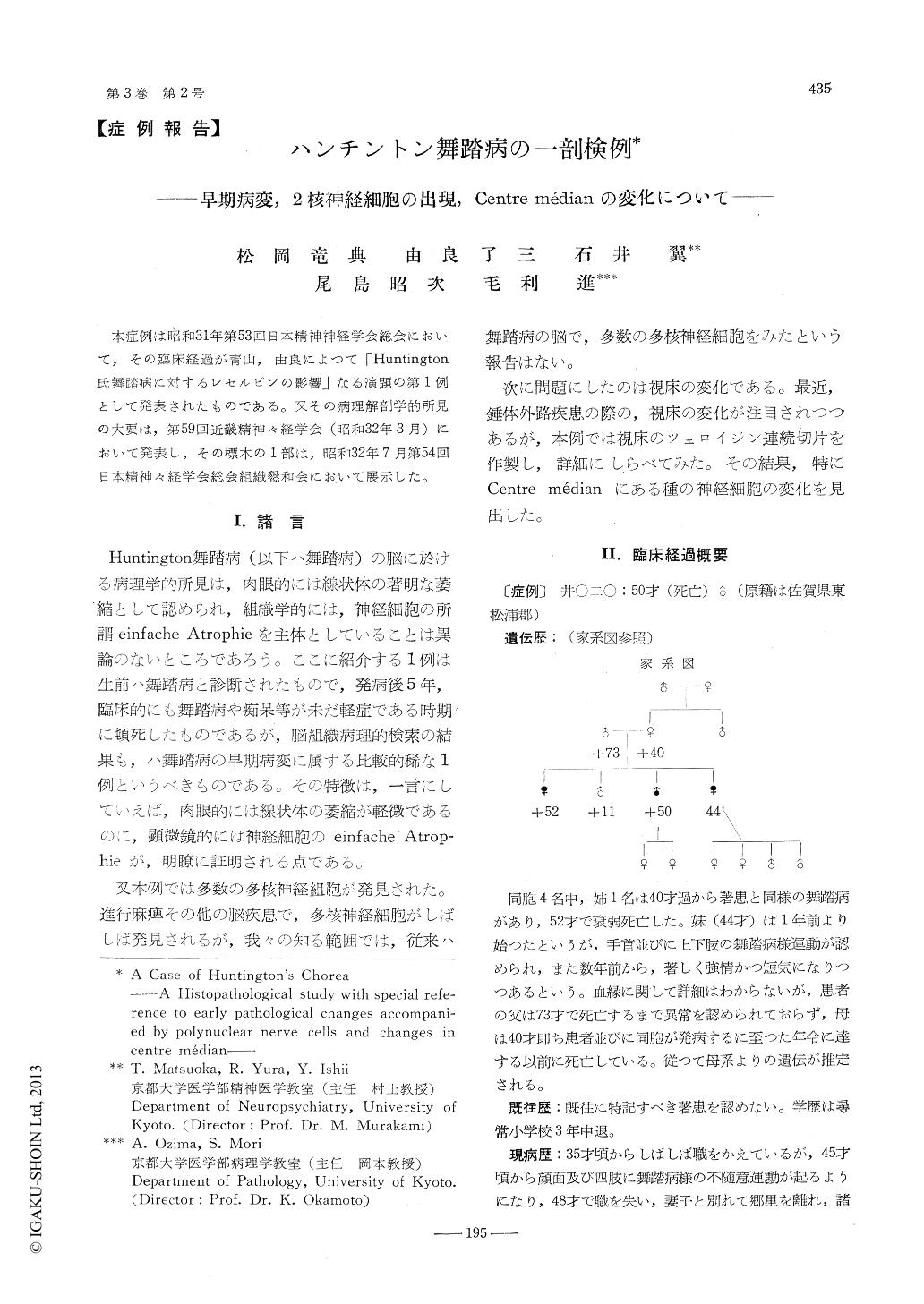
1)遺伝的臨床的に典型的なHuntington舞踏病の1例について組織学的に検索した。臨床経過5年の比較的早期の例である。
2)線状体の萎縮ならびに小神経細胞の脱落は軽微であるが神経細胞の病変は所謂単純萎縮に属するものでありグリアの病変もHuntington舞踏病の典型的な所見に一致する。故に本例の病像はHuntington舞踏病の早期の病変に属するものと考える。
3)線状体以外に皮質,Luis氏体,黒核赤部,視床,視床下灰白質,歯状核,オリーブ核等に病変を見出した。
4)多核神経細胞及び神経細胞の接着傾向を見出した。これらは病変の強い部分に多い。
5)視床のCentre médianに神経細胞の染色性の低下,空胞形成,原形質の融解等を認めた。
6)以上の諸点ならびに萎縮中心の問題,Bindearmchoreaとの関係等に関して考察を行つた。
A male patient, 49-year-old, was admitted toour clinic on account of progressive mental deterioration combined with choreiform movementsin the muscles of the face, extremities, and body, which had started at the age of 44.
He had 4 siblings, 2 of which had been suffering from the same disease. He was supposedto have inherited the disposition from his mother who had died early. When hospitalized, herevealed some changes of character such as irritability, unstableness etc.
His choreiform movements ceased during thefirst reserpine cure, but unfortunately he diedsuddenly from collapse during the second reserpine cure. (at the age of 50)
Autopsy findings. The right ventricle is dilatedand the left ventricle hypertrophied. There areeffusions in the intrathoracic and intraabdominalcavities. The lungs, liver, spleen and kidneysare congested. When the skull is opened, plentyof cerebrospinal fluid runs off. Brain Weight is1260 gr. The lateral ventricles are dilated. Macroscopically atrophy of the cerebral cortex andthe corpus striatum is very slight.
Histological findings. In the corpus striatumsmall ganglion cells are slightly reduced in number, but most of them exhibit remarkable degenerative changes : reduced stainability, swelling of nuclei and protoplasm, liquefaction, vacuole formation, and even pycnotic naked nucleiin the most damaged parts. The large ganglioncells are less affected. The glia manifests reactive phenomena: proliferation and hypertrophy of macro and oligodendroglia, appearance of green-blue granules by Nissl stain in the protoplasmof macroglias, showing positive iron reaction. Similar degenerations of ganglion cells are present in cortex. But glia reactions are slight. The degenerations of ganglion cells are also found in the nucleus dentatus, the corpus Luysi, the thalamus, the hypothalamic nuclei and thenycleus olivae. From the histopathological pointof view, the above mentioned changes supportthe clinical diagnosis of this case, though theatrophy and diminution of the number of ganglion cells are not apparent. The latter fact maybe due to the early histological changes of thisdisease.
Other than typical histological changes of Huntington's chorea we find some interesting changes. Namely, many polynuclear nerve cells arefound in the cortex and the corpus striatum. Most of them are double nucleated and selpomtriple nucleated. They are scattered in the regions where the degenerative changegs are remarkable.
In the centre median of thalamus, we find theganglion cell degenerations without gliareactions,that is, reduced stainability, swelling and liquefaction, vacuole formation and naked nuclei.

Copyright © 1959, Igaku-Shoin Ltd. All rights reserved.


