Japanese
English
- 有料閲覧
- Abstract 文献概要
- 1ページ目 Look Inside
はじめに
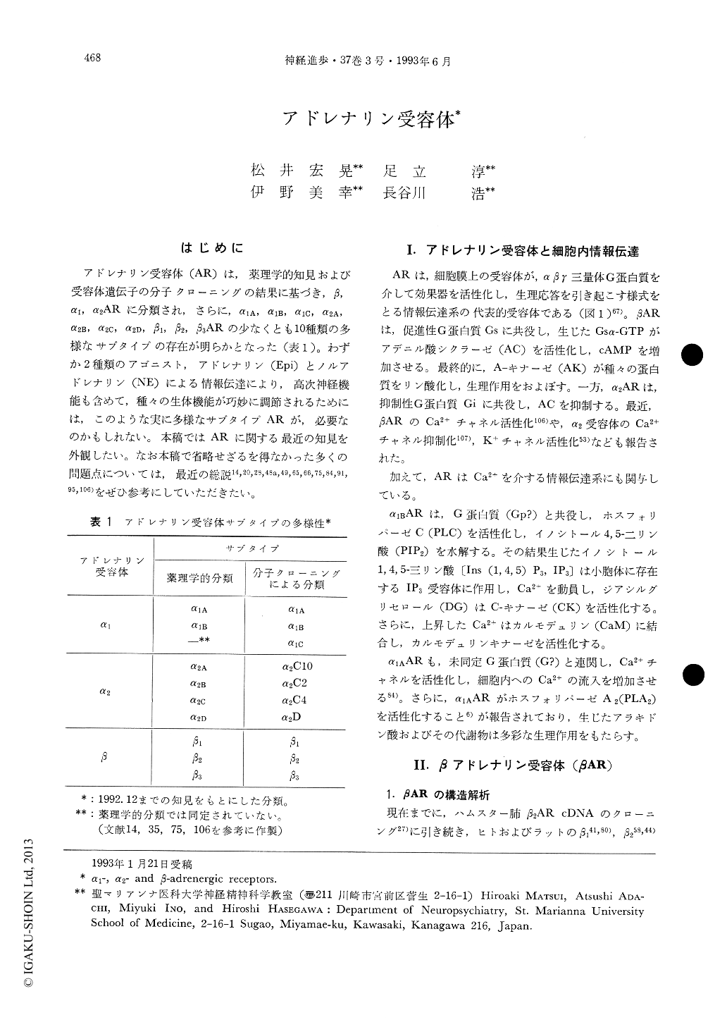
アドレナリン受容体(AR)は,薬理学的知見および受容体遺伝子の分子クローニングの結果に基づき,β,α1,α2ARに分類され,さらに,α1A,α1B,α1C,α2A,α2B,α2C,α2D,β1,β2,β3ARの少なくとも10種類の多様なサブタイプの存在が明らかとなった(表1)。わずか2種類のアゴニスト,アドレナリン(Epi)とノルアドレナリン(NE)による情報伝達により,高次神経機能も含めて,種々の生体機能が巧妙に調節されるためには,このような実に多様なサブタイプARが,必要なのかもしれない。本稿ではARに関する最近の知見を外観したい。なお本稿で省略せざるを得なかった多くの問題点については,最近の総説14,20,28,48a,49,65,66,75,84,91,95,106)をぜひ参考にしていただきたい。
The α1-, α2- and β-adrenergic receptors are member of the superfamily of integral membrane proteins which exert the effects through the interaction with guanine nucleotide-binding regulatory proteins, G proteins. α1-Adrenergic receptors are linked to phosphoinositide-specific phosphodiesterase by Gp, α2-adrenergic receptors to adenylate cyclase by Gi, and β-adrenergic receptor to adenylate cyclase by Gs. From molecular characterization of the adrenergic receptors, it has become evident that this family includes at least ten distinct subtypes. All members of this protein family have the typical features as seven-transmembrane-segment receptors. At least three distinct subtypes exist for each of the β (β1, β2, β3)- and al (α1A, α1B, α1C)-adrenergic receptors, and four subtypes for α2 (α2A, α2B, α2C, α2D)-adrenergic receptors. Each subtypes is present on a distinct chromosome and shows distinct patterns of tissue expression and pharmacological characteristics. Structure and function studies with these wild type and mutated adrenergic receptors have indicated that the ligand binding site is localized within the transmembrane regions, whereas intracellular domains, such as the carboxyl terminal part of the third intracellular loop and the amino terminal part of the carboxyl tail are involved in determining the G protein coupling. Receptor phosphorylation by multiple kinases, such as A kinase and receptor specific kinase, β-adrenergic receptor kinase, appears to be very critical event in regulation of the receptor function in vivo.

Copyright © 1993, Igaku-Shoin Ltd. All rights reserved.


