Japanese
English
- 有料閲覧
- Abstract 文献概要
- 1ページ目 Look Inside
はじめに
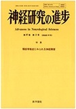
頚椎・頚髄・神経根のおのおのについての解剖学の概略を述べる。しかし,限られた誌面の制約内での概説なので,不明な点や詳細は文献や解剖学の教科書などで確認してほしい。
An anatomical outline of the cervical vertebrae, cervical spinal cord and its nerve roots were described in relation to cervical spondylosis.
First of all, the cervical part of the vertebral column is composed of the vertebrae between which are intervertebral discs. Seven cervical vertebrae form a cervical spinal canal to join the seven verte-bral foramina formed by the vertebral body and arch. Several ligaments support the cervical vertebrae : The anterior and posterior longitudinal ligaments run in front and back of the vertebral body. The ligamentum flavum stretches between the adjacent vertebral arches. Among seven cervical vertebrae, the first and second vertebrae have become modified and are named the atlas and the axis respectively. The main reason of this modification is considered that the atlas supports the skull, and lost its verte-bral body to become the prominent process, the dens of the axis. The combination of the atlas and the axis produces a mechanism for the rotation of the head. Several specialized ligaments to support the mechanism is briefly described. The seventh cervical vertebra which is called the vertebra pro-minens is distinctive because of its long spinous process. The intervertebral foramen through which ventral and dorsal nerve roots, artery and vein pass, is formed between the upper and lower vertebral arches. The measured data of various sizes and angles of the cervical vertebrae were introduced in comparison with those by different methods such as dissecting, X-ray and CT measurements.
Copyright © 1993, Igaku-Shoin Ltd. All rights reserved.