Japanese
English
- 有料閲覧
- Abstract 文献概要
- 1ページ目 Look Inside
- 参考文献 Reference
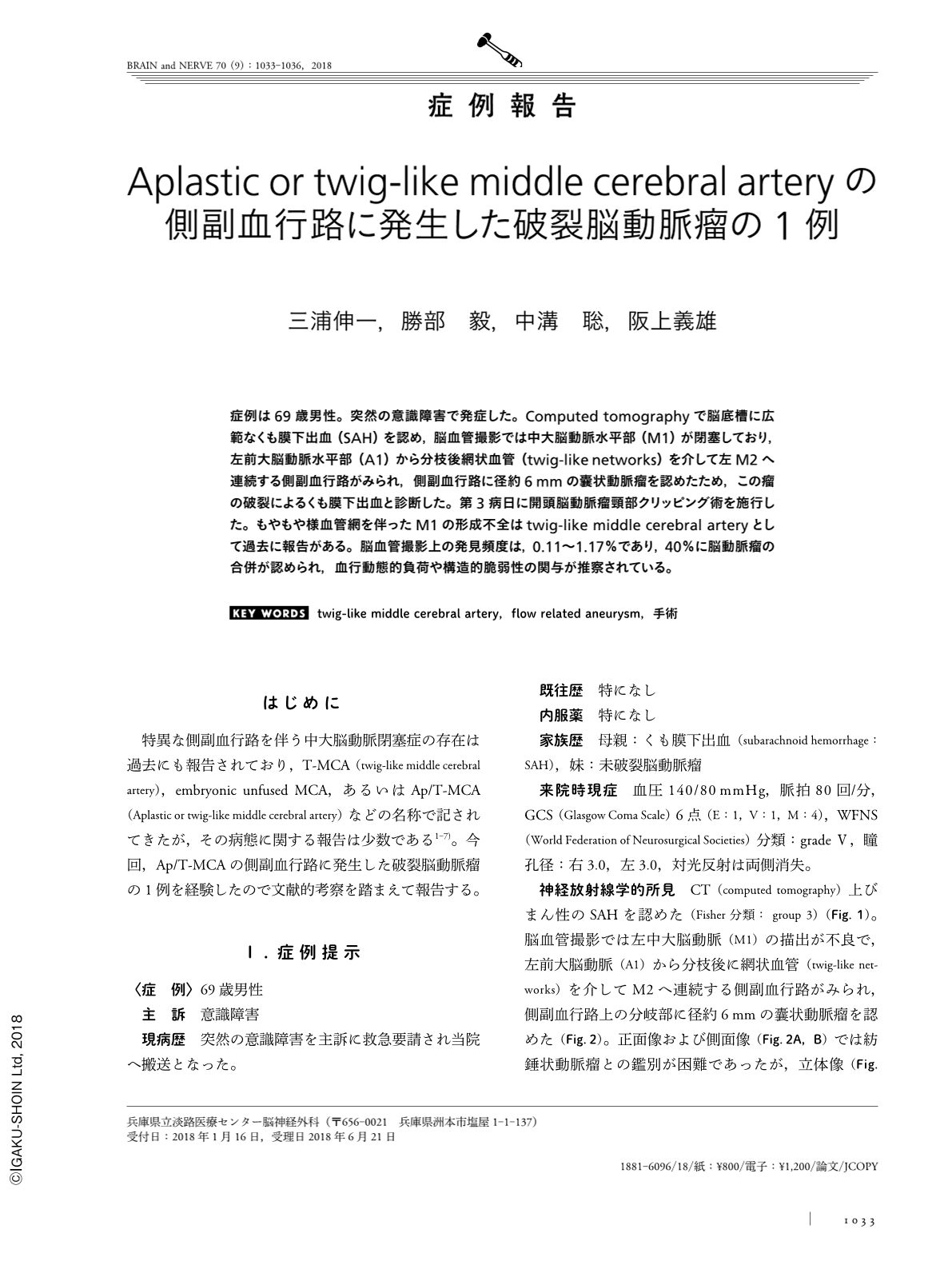
症例は69歳男性。突然の意識障害で発症した。Computed tomographyで脳底槽に広範なくも膜下出血(SAH)を認め,脳血管撮影では中大脳動脈水平部(M1)が閉塞しており,左前大脳動脈水平部(A1)から分枝後網状血管(twig-like networks)を介して左M2へ連続する側副血行路がみられ,側副血行路に径約6mmの囊状動脈瘤を認めたため,この瘤の破裂によるくも膜下出血と診断した。第3病日に開頭脳動脈瘤頸部クリッピング術を施行した。もやもや様血管網を伴ったM1の形成不全はtwig-like middle cerebral arteryとして過去に報告がある。脳血管撮影上の発見頻度は,0.11〜1.17%であり,40%に脳動脈瘤の合併が認められ,血行動態的負荷や構造的脆弱性の関与が推察されている。
Abstract
A 69-year-old man presented with sudden loss of consciousness and was admitted to our hospital. Computed tomography revealed diffuse subarachnoid hemorrhage. Digital subtraction angiography revealed occlusion of the left M1 segment, collateral arteries from the left A1 to the left M2 via twig-like networks, and a 6-mm aneurysm in the collateral arteries. Clipping surgery was performed on the 3rd hospital day. Vascular abnormalities of the middle cerebral artery with twig-like networks have been reported with an incidence of 0.11-0.17%. In addition, aneurysms are reported as complications in 40% of cases, suggesting hemodynamic stress and structural vulnerability.
(Received January 16, 2018; Accepted June 21, 2018; Published September 1, 2018)

Copyright © 2018, Igaku-Shoin Ltd. All rights reserved.


