Japanese
English
- 有料閲覧
- Abstract 文献概要
- 1ページ目 Look Inside
I.はじめに
Neurofibromatosis(以下NF)は遺伝性の中胚葉および外胚葉系の形成異常をきたす神経皮膚症候群のひとつで21),多発性神経線維腫,骨病変が主症状であるが,時に血管病変を合併することが知られている5).NFに合併する頭蓋内血管病変は,内頸動脈系の閉塞2,8-10,12,17,19,22)およびモヤモヤ血管の形成や動脈瘤発生2,13,19)が報告され,その機序として血管壁内のSchwann細胞の増殖やそれに伴う2次的な変性が推定されている16).最近われわれは,右内頸動脈の狭窄と,側副血行路として発達したHeubner's arteryの末梢部に破裂脳動脈瘤を合併したNFのまれな1例を経験したので若干の文献的考察を加えて報告する.
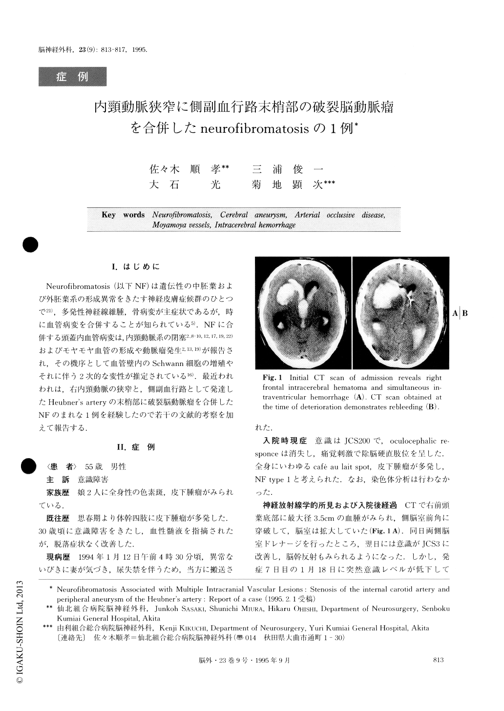
Neurofibromatosis (NF), or von Recklinghausen's disease, is an autosomal dominant disorder of both mesoderm and ectoderm dysplasia commonly character-ized by numerous neurofibromas and cafe-au-lait spots. However, cerebrovascular diseases associated with NF are rare, and among them occlusive vascular lesions have already been reported in the literature. It is postu-lated that the pathogenesis of the associated vascular lesions may be attributable to the proliferation of Schwann cells and the subsequent degeneration in the vessel wall. We documented here an uncommon case of NF associated with massive intracerebral hemorrhage caused by an aneurysm at the distal portion of the di-lated Heubner's artery. Intracranial hemorrhage in asso-ciation with NF is reviewed in the literature, and the clinical profiles, type and origin of bleeding, and associ-ated occlusive vascular lesions are discussed. A 55-year-old man was admitted to our hospital on January 12, 1994, after he had suddenly become com-atose. The patient was known to be affected by NF from both the family and past histories. On admission, the patient demonstrated a decerebrate posture in re-sponse to painful stimuli, and was then graded as 200 according to the Japan Coma Scale (JCS). CT scan showed a significant amount of blood clots present at the base of the right frontal lobe and in the lateral and third ventricles. After bilateral ventriculostomies were carried out for continuous drainage of bloody cerebro-spinal fluid, the patient continued to regain partial con-sciousness to the level of JCS grade 3 until the seventh day, when he again suddenly deteriorated and became comatose. Emergency CT confirmed an evidence of re-bleeding. Right carotid angiograms demonstrated marked stenosis of the right internal carotid artery dis-tal to the junction of the anterior choroidal artery, and the right middle cerebral arteries were faintly opacified with abnormal vascular network (moyamoya vessels). Left carotid angiograms revealed cross circulation to the contralateral hemisphere through the anterior com-municating artery. The right Heubner's artery was found dilated and directly connected to the right middle cerebral artery, and two saccular aneurysms were seen at the peripheral portion of the artery. At surgery a lar-ger aneurysm, presumed responsible for the current bleeding, was clipped in an angioplastic fashion by two Sugita straight clips by means of a transcortical approach through the hematoma cavity. Postoperative angiograms showed the disappearance of both the aneurysms.

Copyright © 1995, Igaku-Shoin Ltd. All rights reserved.


