Japanese
English
- 有料閲覧
- Abstract 文献概要
- 1ページ目 Look Inside
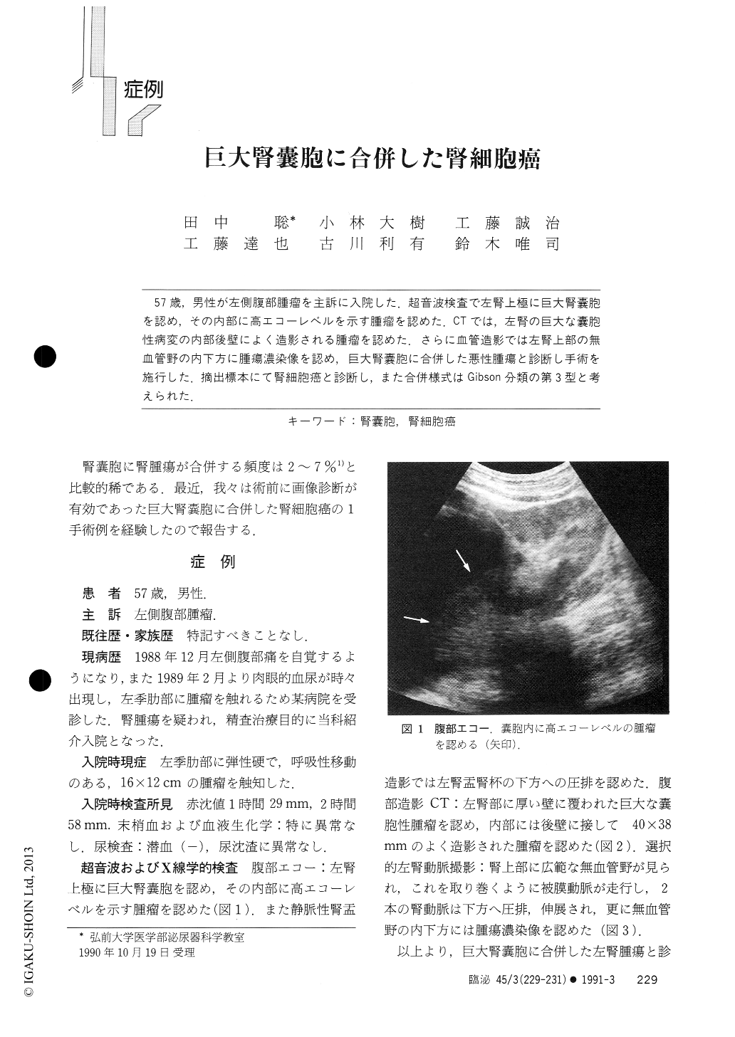
57歳,男性が左側腹部腫瘤を主訴に入院した.超音波検査で左腎上極に巨大腎嚢胞を認め,その内部に高エコーレベルを示す腫瘤を認めた.CTでは,左腎の巨大な嚢胞性病変の内部後壁によく造影される腫瘤を認めた.さらに血管造影では左腎上部の無血管野の内下方に腫瘍濃染像を認め,巨大腎嚢胞に合併した悪性腫瘍と診断し手術を施行した.摘出標本にて腎細胞癌と診断し,また合併様式はGibson分類の第3型と考えられた.
A 57-year-old man was admitted with a chief complaint of a left flank mass. Ultrasonography demon-strated a high echoic mass lesion in a giant renal cyst in upper portion of the left kidney. Contrast enhanced computed tomography revealed a well-enhanced lesion on the posterior wall of the giant cyst in the left kidney, and selective renal angiography indicated tumor staining at the inner posterior portion of the avascular area of the left kidney. Diagnosis of a giant renal cyst with malignancy was made, and a left nephrectomy was performed.

Copyright © 1991, Igaku-Shoin Ltd. All rights reserved.


