Japanese
English
- 有料閲覧
- Abstract 文献概要
- 1ページ目 Look Inside
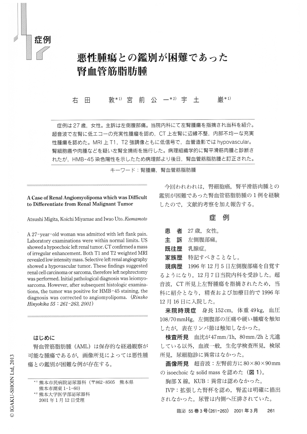
症例は27歳,女性。主訴は左側腹部痛。当院内科にて左腎腫瘍を指摘され当科を紹介。超音波で左腎に低エコーの充実性腫瘤を認め,CT上左腎に辺縁不整,内部不均一な充実性腫瘍を認めた。MRI上T1,T2強調像ともに低信号で,血管造影ではhypovascular。腎細胞癌や肉腫などを疑い左腎全摘術を施行した。病理組織学的に腎平滑筋肉腫と診断されたが,HMB−45染色陽性を示したため病理部より後日,腎血管筋脂肪腫と訂正された。
A 27-year-old woman was admitted with left flank pail. Laboratory examinations were within normal limits. U showed a hypoechoic left renal tumor. CT confirmed a mass of irregular enhancement. Both Ti and T2 weighted MRI revealed low intensity mass. Selective left renal angiography showed a hypovascular tumor. These findings suggestedrenal cell carcinoma or sarcoma, therefore left nephrectomy was performed. Initial pathological diagnosis was leiomyo-sarcoma. However, after subsequent histologic examina tions, the tumor was positive for HMB-45 staining, the diagnosis was corrected to angiomyolipoma.

Copyright © 2001, Igaku-Shoin Ltd. All rights reserved.


