Japanese
English
- 有料閲覧
- Abstract 文献概要
- 1ページ目 Look Inside
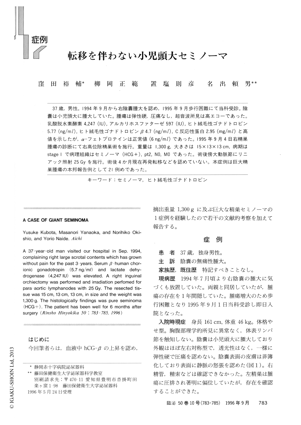
37歳,男性。1994年9月から右陰嚢腫大を認め,1995年9月歩行困難にて当科受診。陰嚢は小児頭大に腫大していた。腫瘍は弾性硬,圧痛なし,超音波所見は高エコーであった。乳酸脱水素酵素4,247(IU),アルカリホスファターゼ597(IU),ヒト絨毛性ゴナドトロピン5.77(ng/ml),ヒト絨毛性ゴナドトロピンβ4.7(ng/ml),C反応性蛋白2.95(mg/ml)と高値を示したが,α-フェトプロテインは正常値(6ng/ml)であった。1995年9月4日右精巣腫瘍の診断にて右高位除精巣術を施行。重量は1,300g,大きさは15×13×13cm,病期はstage 1で病理組織はセミノーマ(HCG+),pt2, NO, MOであった。術後傍大動脈節にリニアック照射25Gyを施行。術後4か月現在再発転移などを認めていない。本症例は巨大精巣腫瘍の本邦報告例として21例めであった。
A 37-year-old man visited our hospital in Sep. 1994, complaining right large scrotal contents which has grown without pain for the past 3-years. Serum β-human chorionic gonadotropin (5.7ng/ml) and lactate dehydrogenase (4,247IU) was elevated. A right inguinal orchiectomy was performed and irradiation perfomed for para-aortic-lymphanodes with 25Gy. The resected tissue was 15cm, 13cm, 13cm, in size and the weight was 1,300g. The histologically findings was pure seminoma (HCG+). The patient has been well for 6 months after surgery

Copyright © 1996, Igaku-Shoin Ltd. All rights reserved.


