Japanese
English
- 有料閲覧
- Abstract 文献概要
- 1ページ目 Look Inside
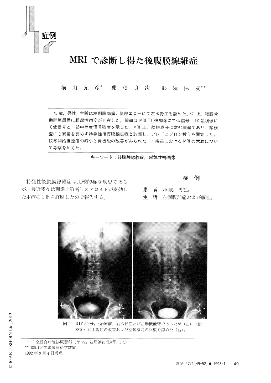
75歳,男性。主訴は左側腹部痛。腹部エコーにて左水腎症を認めた。CT上,総腸骨動静脈周囲に腫瘤性病変が存在した。腫瘤はMRI T1強調像にて低信号,T2強調像にて低信号と一部中等度信号強度を示した。MRI上,線維成分に富む腫瘤であり,諸検査にも異常を認めず特発性後腹膜線維症と診断し,プレドニゾロン投与を開始した。投与開始後腫瘤の縮小と腎機能の改善がみられた。本疾患におけるMRIの意義について考察を加えた。
The patient was a 75-year-old man who complained of left flank pain showed left hydronephrosis by ultrasound sonography. Computed tomography (CT) proved mass lesion around bilateral iliac arteries. In magnetic resonance imaging (MRI), they appeared at low intensity on T1 weighted image and at intermedi-ate intensity on T2 weighted image. which suggested fibrous component of the mass. He was diagnosed as retroperitoneal fibrosis and treated by prednisolone alone. Following the therapy, the mass had reduced. We considered that MRI was very useful method for diagnosis, and follow up of idiopathic retroperitoneal fibrosis.

Copyright © 1993, Igaku-Shoin Ltd. All rights reserved.


