Japanese
English
- 有料閲覧
- Abstract 文献概要
- 1ページ目 Look Inside
Lichen sclerosus et atrophicusは比較的まれな疾患ではあるが,その臨床像に特異な所見を有するにかかわらず,1887年のHallopeau1)の最初の報告以来その独立性に関して種々論議がなされ,ことに扁平苔癬あるいはMorpheaとの異同については臨床像,主として組織学的所見を中心に数多の記載がある。しかしKogoj2)らによつて扁平苔癬から分離され,Montgornery & Hi-ll3),Miescher4)らの臨床的組織学的検索から本症の独立性が支持され,以後独立疾患とするものが多く本症の概念はほぼ明瞭になつてきている。しかし初期像に関してはいまだ意見の一致をみない点が少なくないようである。
一方,本邦においては小堀ら5)の報告以来,相次いで症例追加がなされ10例あまりが数えられ,本症の臨床像はようやく明確にされてきた。最近,舌変化を伴い全身に対側性に皮疹の発生をみた本症の1例を経験し,初期像ならびに組織化学的に2・3検討を加えたので報告する。
A 42-year-old man noticed an erythematoatrophic lesion on the left surface of the tongue about 9 years ago. On the next year he was affected by numerous, itchy, white papules distri-buted symmetrically over the thighs, nape, face, dorsa of the hands, feet and fingers, which enlarged gradually and fused into the large sclerotic patches covered with wavy, tightly ad-hering characteristic scales. They have become the white, scar-like, atrophic, depressing patches gradually. One year ago, numerous, white papules with comedo-like, horny plugging on the center and surrounded by slight erythematous area, were appeared symmetrically on the fore-arms. At times vesicles appeared on the old lesions on the thighs and dorsa of the feet, which reptured and left intractable ulcers. Nails of the fingers and toes were atrophic, and some of them have fallen off.
Histologic specimen from the early lesion of the forearm showed the marked hyperkeratosis with horny plugging, narrow edematous zone of the dermis without elastic fibers, just beneath the epidermis, mild atrophy of the epidermis, a mild hydropic degeneration of the basal cell layer, and slight sclerosis of the dermis.
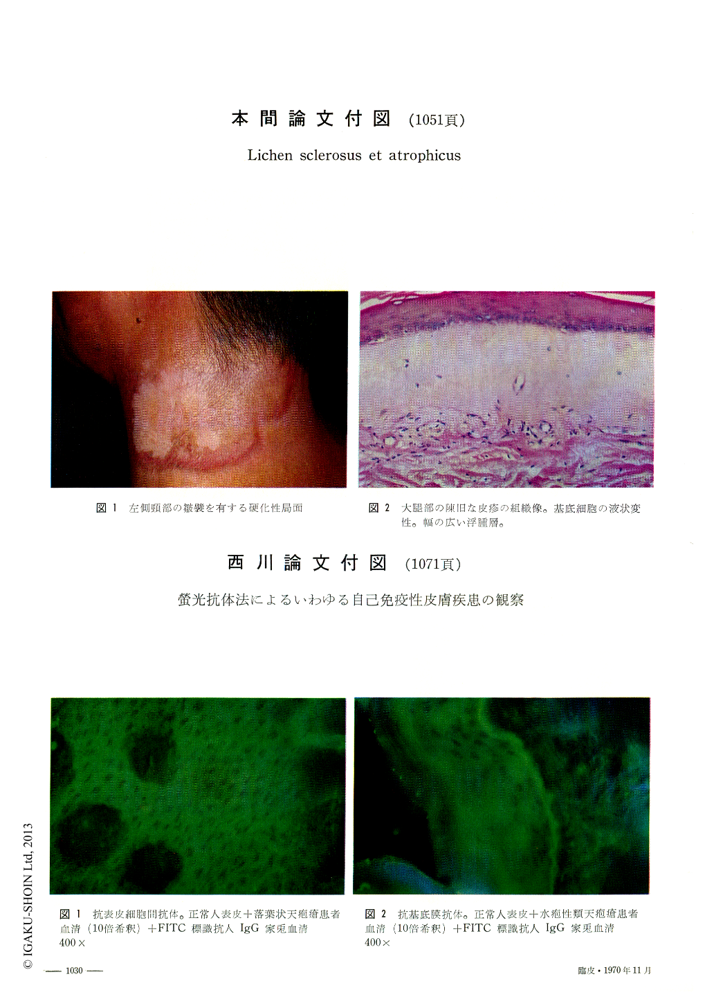
Those from the old lesions on the thigh showed marked hyperkeratosis, wide edematous zone in the upperdermis, marked atrophy of the epidermis, severe hydropic degeneration of the basal cells, and sclerotic changes of the dermis accompanied with proliferation of the elastic fibers, the dendritic branches of which invaded into the edematous zone.
Histochemical analysis revealed that the edematous zone was occupied with the fluid of the high molecular, fibrous protein, which might have induced proliferation of collagenous and elastic fibers secondarily.

Copyright © 1970, Igaku-Shoin Ltd. All rights reserved.


