Japanese
English
- 有料閲覧
- Abstract 文献概要
- 1ページ目 Look Inside
従来クリプトコックス症としては髄膜炎の形で発症する場合が多いとされていたが,近年肺,肝,脾,腎,リンパ腺,などという各臓器の罹患例の報告もかなり散見される。もつとも他臓器に比し本症が皮膚に発症したという例はなお依然として少ない。しかもこれまでの皮膚クリプトコックス症症例のうちには,皮膚以外の病巣からの全身播種の1部として皮膚に発症した例,すなわち続発性皮膚クリプトコックス症と考えられる症例が多く,原発性皮膚クリプトコックス症はきわめて少ない。
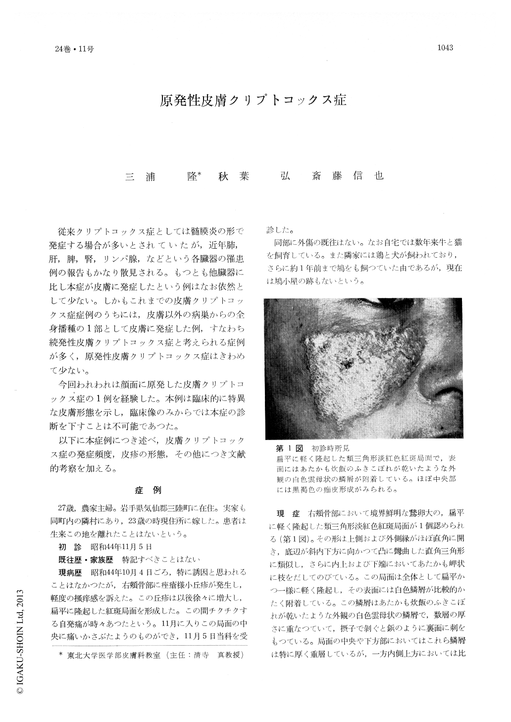
今回われわれは顔面に原発した皮膚クリプトコックス症の1例を経験した。本例は臨床的に特異な皮膚形態を示し,臨床像のみからでは本症の診断を下すことは不可能であつた。
A case of this disease in a 27-year-old farmer's wife living in Kese county of Iwate prefecture was reported. The skin eruption was the triangular erythematous patch covered with the thick, white lamellar, micaceous scales, resembling to the discoid 1.e. on the right malar eminence.
The results of the laboratory tests were within the normal limits.
The histological specimen showed the chronic granulomatous reaction with many spores which existed freely in the tissue or inside the giant cells. The fungi were identified as a crypto-coccis neoformans. The lesion disappeared after the continuous, intravenous irrigation of the total dose of 600 mg of Amphotericin B for 100 days.

Copyright © 1970, Igaku-Shoin Ltd. All rights reserved.


