Japanese
English
- 有料閲覧
- Abstract 文献概要
- 1ページ目 Look Inside
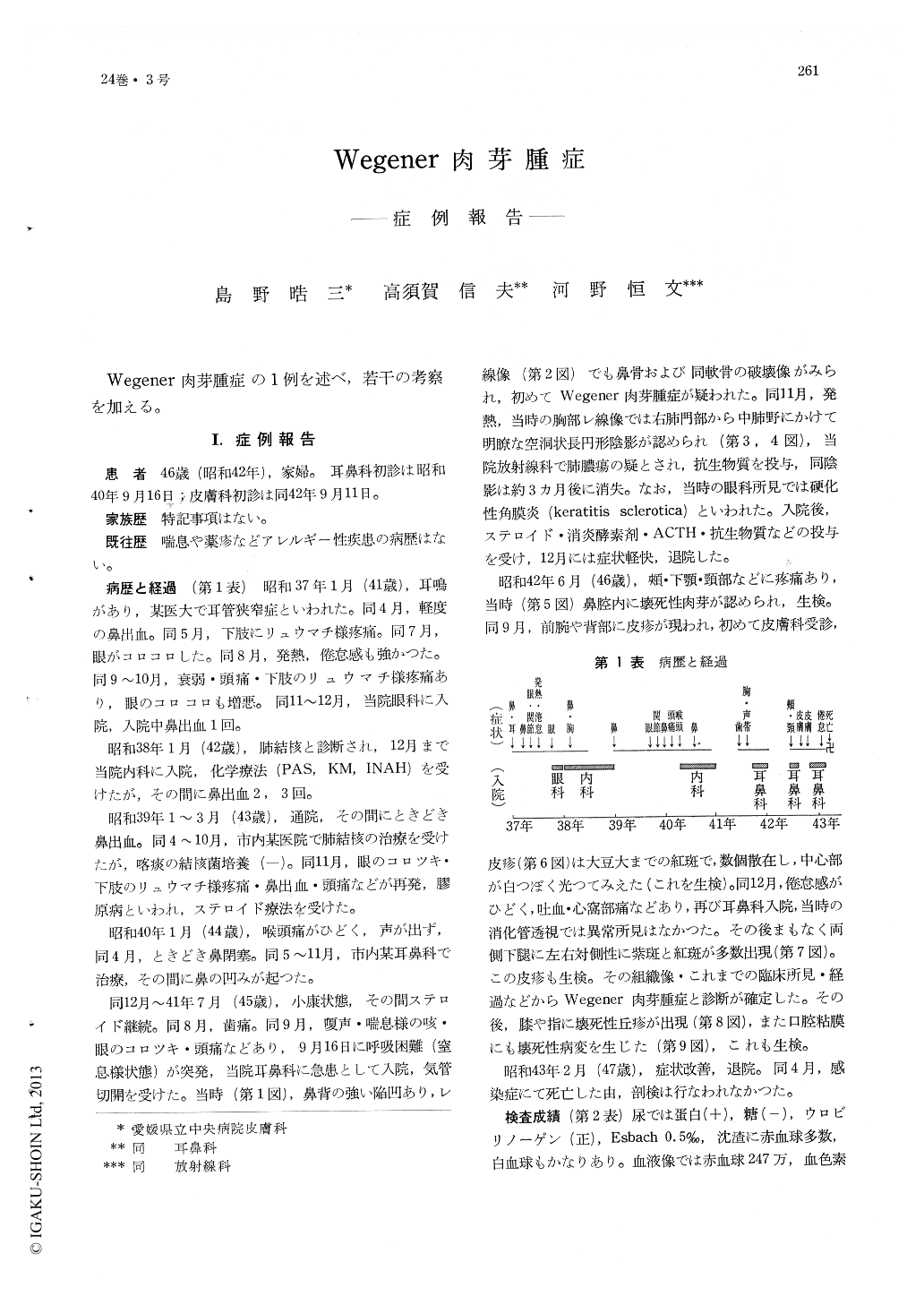
Wegener肉芽腫症の1例を述べ,若干の考察を加える。
A 46-year-old housewife visited the author's clinic on September 11, 1967. Past history did not disclose bronchial asthma or drug allergy. Her disease started in January 1962 with epistaxis and buzzing in the ears. Thereafter various symptoms such as arthralgia, fever, weakness, headache, hoarseness, etc. appeared and disappeared. She was variously diagnosed as having stenosis of the eustachian canal, a cold, pulmonary tuberculosis or a collagenous disease. She was admitted to E. N. T. Clinic of the Hospital under asphyxia in September 1966, and received an emergency tracheotomy. At that time she had a suddle nose, In October a roentgenogram of the chest revealed a round shadow in the right lung, but Mycobacterium tuberculosis was negative in the sputum. In June 1967 necrotizing foci in the nasal cavity and in September 1967 a disseminated erythemato-purpuric eruption were noticed. After that time, necrotizing papules on the knees and fingers and large necrotizing lesions on the buccal and gingival mucous membrane appeared.
In Februay 1968, she was discharged and died of some infection in April 1968. Autopsy wuas not be performed. Laboratory tests revealed severe anemia, accelerated ESR, albuminuria, hypo-proteinemia, moderatly increased urea nitrogen and 1 plus CRP.
Histologic specimen from a necrotizing lesion in the nasal cavity showed necrotic foci with nuclear fragments surrounded by a dense infiltrate composed mainly of polymorphonuclear cells accompanied by plasma cells, histiocytes, eosinophils and giant cells.
Histologic specimen from an erythemato-purpuric lesion on the lower leg showed necrotizing angitis with fibrinoid degeneration, nuclear fragments, polymorphonuclear cells and eosinophils surrounded by an infiltrated zone of histiocytes and giant cells.
Specimen from a necrotic lesion in the oral cavity showed almost the same picture as that of the necrotic lesion in the nasal cavity.
Entire course of the patient was 6 years and 4 months, and peroral administration of cortico-steroids was effective on suppression of the symptoms.

Copyright © 1970, Igaku-Shoin Ltd. All rights reserved.


