Japanese
English
- 有料閲覧
- Abstract 文献概要
- 1ページ目 Look Inside
緒言
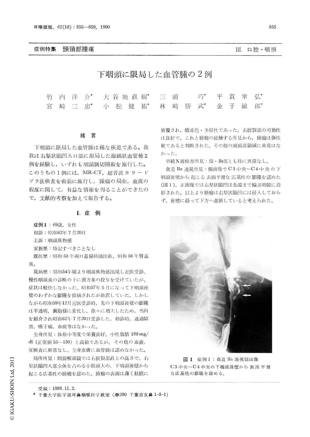
下咽頭に限局した血管腫は稀な疾患である。我我は右梨状陥凹入口部に限局した海綿状血管種2例を経験し,いずれも咽頭側切開術を施行した。このうちの1例には,MR-CT,超音波カラードプラ法検査を術前に施行し,腫瘤の局在,血流の程度に関して,有益な情報を得ることができたので,文献的考察を加えて報告する。
We reported two cases of hypopharyngeal cavernous hemangiomas in a 69-year-old and a 43-year-old female. Both of them showed a localized lesion in right piryform sinus. Both were treated by lateral pharyngotomy without tracheostomy, and had good prognosis. MR-CT and the color doppler imaging were used in the preoperative study of the first case. MR-CT imaging was use-ful for determining localization of hemangioma, and color doppler imaging was also useful for the observation of blood flow inside hemangioma. We could not detect the blood flow signals in the first case, and therefore, it was not necessary to perform angiography.

Copyright © 1990, Igaku-Shoin Ltd. All rights reserved.


