Japanese
English
- 有料閲覧
- Abstract 文献概要
- 1ページ目 Look Inside
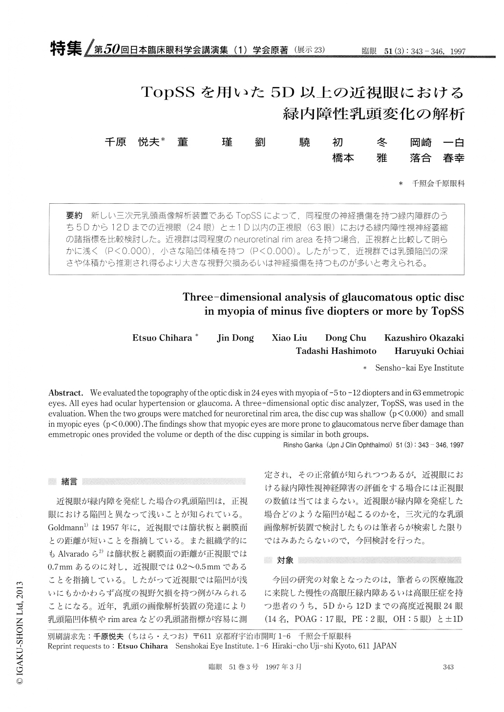
(展示23) 新しい三次元乳頭画像解析装置であるTopSSによって,同程度の神経損傷を持つ緑内障群のうち5Dから12Dまでの近視眼(24眼)と±1D以内の正視眼(63眼)における緑内障性視神経萎縮の諸指標を比較検討した。近視群は同程度のneuroretinal rim areaを持つ場合,正視群と比較して明らかに浅く(P<0.000),小さな陥凹体積を持つ(P<0.000)。したがって,近視群では乳頭陥凹の深さや体積から推測され得るより大きな視野欠損あるいは神経損傷を持つものが多いと考えられる。
We evaluated the topography of the optic disk in 24 eyes with myopia of -5 to -12 diopters and in 63 emmetropic eyes. All eyes had ocular hypertension or glaucoma. A three-dimensional optic disc analyzer, TopSS, was used in the evaluation. When the two groups were matched for neuroretinal rim area, the disc cup was shallow (p < 0.000) and small in myopic eyes (p <0.000) .The findings show that myopic eyes are more prone to glaucomatous nerve fiber damage than emmetropic ones provided the volume or depth of the disc cupping is similar in both groups.

Copyright © 1997, Igaku-Shoin Ltd. All rights reserved.


