Japanese
English
- 有料閲覧
- Abstract 文献概要
- 1ページ目 Look Inside
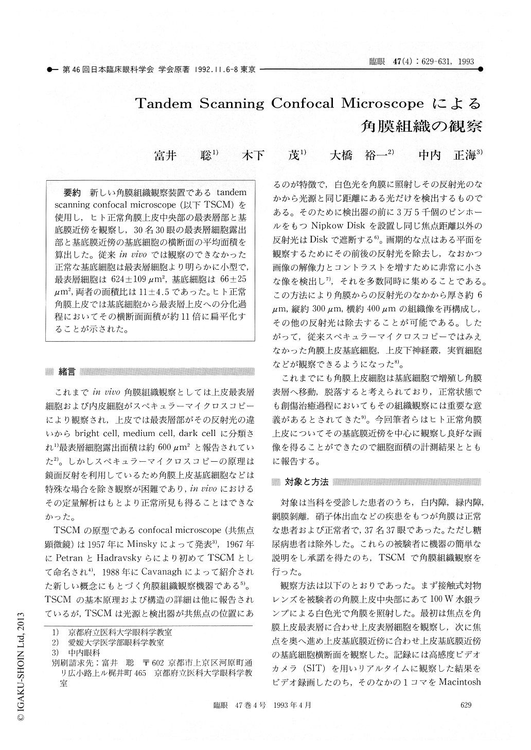
新しい角膜組織観察装置であるtandemscanning confocal microscope (以下TSCM)を使用し,ヒト正常角膜上皮中央部の最表層部と基底膜近傍を観察し,30名30眼の最表層細胞露出部と基底膜近傍の基底細胞の横断面の平均面積を算出した。従来in vivoでは観察のできなかった正常な基底細胞は最表層細胞より明らかに小型で,最表層細胞は624±109μm2,基底細胞は66±25μm2,両者の面積比は11±4.5であった。ヒト正常角膜上皮では基底細胞から最表層上皮への分化過程においてその横断面面積が約11倍に扁平化することが示された。
The tandem scanning confocal microscope, TSCM, allows an in vivo observation of corneal epithelium, nerve fibers and endothelial cells. We applied this method to 30 healthy eyes in 30 persons aged from 22 to 90 years. Slitlamp findings were normal in all the eyes. The findings of the most superficial cells and basal cells close to the basallamina in the central cornea were recorded on videotape and were analyzed by a computer-assist-ed digitizer. The exposed area of the most superfi-cial epithelial cells was 624±109 μm2 and the area at the horizontal section of basal cells was 66±4.5 μm2. The ratio of area of superficial cells to basal cells was thus 11.0±4.5. TSCM was thus useful in evaluating the relationship between superficial and basal cells in normal human cornea in vivo.

Copyright © 1993, Igaku-Shoin Ltd. All rights reserved.


