Japanese
English
- 有料閲覧
- Abstract 文献概要
- 1ページ目 Look Inside
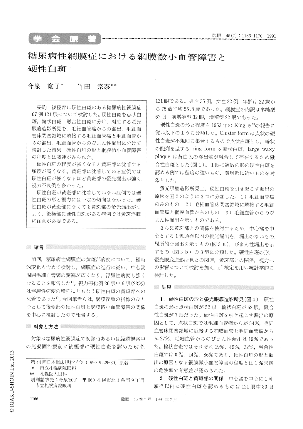
後極部に硬性白斑のある糖尿病性網膜症67例121眼について検討した。硬性白斑を点状白斑,輪状白斑,融合性白斑に分け,対応する螢光眼底造影所見を,毛細血管瘤からの漏出,毛細血管床閉塞領域に隣接する毛細血管瘤と毛細血管からの漏出,毛細血管からのびまん性漏出に分けて検討した結果,硬性白斑の形と網膜微小血管障害の程度とは関連がみられた。
硬性白斑の程度が強くなると黄斑部に沈着する頻度が高くなる。黄斑部に沈着している症例では硬性白斑が強くなるほど黄斑部の螢光漏出が強く,視力不良例も多かった。
硬性白斑が黄斑部に沈着していない症例では硬性白斑の形と視力には一定の傾向はなかった。硬性白斑が黄斑部になくても黄斑部の螢光漏出がつよく,後極部に硬性白斑がある症例では黄斑浮腫に注意が必要である。
We evaluated 121 eyes in 67 patients with diabet-ic retinopathy and hard exudates in the posteriorfundus.The hard exudates appeared as cluster in 52eyes, circinate in 62 and confluent into large waxyplaque in 7.The degree of retinal microangiopathywas divided into 3 groups by fluorescein angiogra-phic findings:those with focal leakage from mi-croaneurysms only, leakage from microaneurysmsand capillaries adjacent to nonperfused areas, anddiffuse leakage from dilated retinal capillaries. Theincidence of each group of microangiopathy was: 54%, 27% and 19% in cluster form, 19%, 49% and32% in circinate one, and 0%, 14% and 86% inwaxy plaque. The types of hard exudates andretinal microangiopathy were significantly cor-related (p<0.01).
Incidence of macular hard exudates was 52% incluster group, 76% in circinate one and 86% inwaxy plaque group. The amount of macular hardexudates was positively correlated with impair-ment of visual acuity.

Copyright © 1991, Igaku-Shoin Ltd. All rights reserved.


