Japanese
English
- 有料閲覧
PPV購入案内
1,320円 (1,200円+税10%) こちらは、1013ページから1016ページの文献です。購入には医書.jp本会員登録が必要です。
会員登録完了後に改めて上記「購入サイトへ」を選択してください。
または「購入サイトへ」選択後に「購入ログイン」、「新規会員登録」の順に進んでください。
会員登録について詳しくはをご確認ください。
- Abstract 文献概要
- 1ページ目 Look Inside
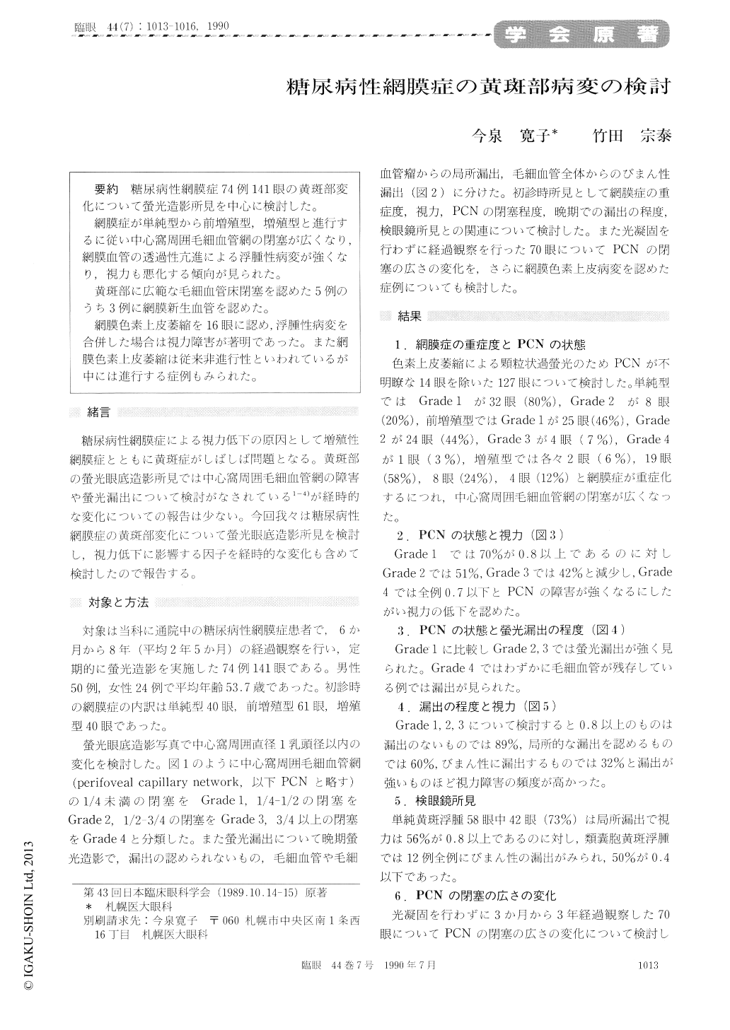
糖尿病性網膜症74例141眼の黄斑部変化について螢光造影所見を中心に検討した。
網膜症が単純型から前増殖型,増殖型と進行するに従い中心窩周囲毛細血管網の閉塞が広くなり,網膜血管の透過性亢進による浮腫性病変が強くなり,視力も悪化する傾向が見られた。
黄斑部に広範な毛細血管床閉塞を認めた5例のうち3例に網膜新生血管を認めた。
網膜色素上皮萎縮を16眼に認め,浮腫性病変を合併した場合は視力障害が著明であった。また網膜色素上皮萎縮は従来非進行性といわれているが中には進行する症例もみられた。
We evaluated the macular lesions in 141 eyes, 74 cases, of diabetic retinopathy. The diabetic retinopathy was simple in 40 eyes, preproliferative in 61 and proliferative in 40. We used fluorescein angiography in the evaluation.
Perifoveal capillary occlusion was significantly associated with increased extravasation of dye in the macula and with impairment of visual acuity. In 3 out of 5 eyes with extensive perifoveal capillarynonperfusion, neovascularization developed from perifoveal retinal vessels. There were 16 eyes show-ing granular background hyperfluorescence sugges-tive of retinal pigment epithelial atrophy. The presence of macular edema was generally associat-ed with poor visual acuity. The retinal pigment atrophy progressed to pattern dystrophy in some eyes.
Copyright © 1990, Igaku-Shoin Ltd. All rights reserved.