Japanese
English
- 有料閲覧
- Abstract 文献概要
- 1ページ目 Look Inside
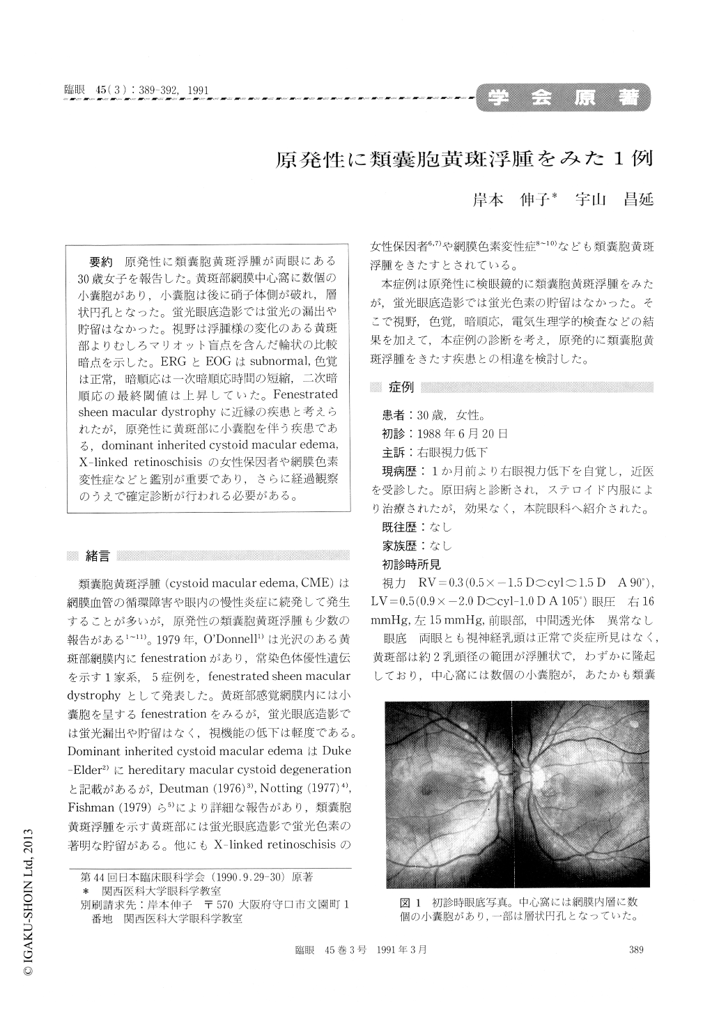
原発性に類嚢胞黄斑浮腫が両眼にある30歳女子を報告した。黄斑部網膜中心窩に数個の小嚢胞があり,小嚢胞は後に硝子体側が破れ,層状円孔となった。蛍光眼底造影では蛍光の漏出や貯留はなかった。視野は浮腫様の変化のある黄斑部よりむしろマリオット盲点を含んだ輪状の比較暗点を示した。ERGとEOGはsubnormal,色覚は正常,暗順応は一次暗順応時間の短縮,二次暗順応の最終閾値は上昇していた。Fenestratedsheen macular dystrophyに近縁の疾患と考えられたが,原発性に黄斑部に小嚢胞を伴う疾患である,dominant inherited cystoid macular edema,X-linked retinoschisisの女性保因者や網膜色素変性症などと鑑別が重要であり,さらに経過観察のうえで確定診断が行われる必要がある。
Dept of Ophthalmol, Kansai Med Univ A 30-year-old woman showed with primary cystoid macular edema. In her central macular area, several cysts were seemred within the sensory retina. The cysts raptured in the vitreous side, and appeared like lamellar hole during the follow up course. Fluorescein angiography showed neither leaking nor pooling of dye. Visual field showed relative ring scotoma including blind spot of Mar-iotte, so the sensitivity more strongly reduced the outer area from the edematous macula. ERG and EOG were subnormal. No abnormality in color vision could be detected. Dark adaptation demon-strated a shortened cone plateau with elevated final rod threshold.
Ophthalmoscopically the fundus of this case was seem as cystoid macular edema, that were Domi-nant inherited cystoid macular edema, but physico-psychiatric examination suggested Fenestrated sheen macular dystrophy, but differential diagnosis was difficult.
So we needed longer term follow-up to confirm the diagnoses.

Copyright © 1991, Igaku-Shoin Ltd. All rights reserved.


