Japanese
English
- 有料閲覧
- Abstract 文献概要
- 1ページ目 Look Inside
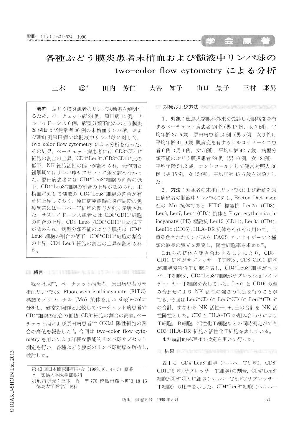
ぶどう膜炎患者のリンパ球動態を解明するため,ベーチェット病24例,原田病14例,サルコイドーシス6例,病型分類不能のぶどう膜炎28例および健常者30例の末梢血リンパ球,および新鮮例原田病では髄液中リンパ球に対して,two-color flow cytometryによる分析を行った。その結果,ベーチェット病患者にはCD8+CD11+細胞の割合の上昇,CD4+Leu8−/CD8+CD11+比の低下,NK細胞活性の低下が認められ,発作期と緩解期ではリンパ球サブセットに差を認めなかった。原田病患者にはCD4+Leu8−細胞の割合の低下,CD4+Leu8+細胞の割合の上昇が認められ,末梢血に対して髄液のCD4+Leu8−細胞の割合が有意に上昇しており,原田病発症時の炎症局所の免疫異常にはヘルパーT細胞の関与が強く示唆された。サスコイドーシス患者にはCD8+CD11+細胞の割合の上昇,CD4+Leu8−/CD8+CD11+比の低下が認められ,病型分類不能のぶどう膜炎はCD4+Leu8−細胞の割合の低下,CD8+CD11+細胞の割合の上昇,CD4+Leu8+細胞の割合の上昇が認められた。
We evaluated the T lymphocyte subsets in the peripheral blood and the cerebrospinal fluid in patients with uveitis. Two-color flow cytometry was used. The cases comprised Behçet's disease with ocular involvement 24 patients, Vogt-Koyanagi-Harada syndrome 14, sarcoidosis 6, unclassified uveitis 28 and 30 healthy volunteers as control. Lymphocytes were stained by fluorescein isothiocyanate labelling anti Leu2a (CD8), Leu8, Leu7 and Leu4 (CD3) antibodies and by phycoer-ythrin labelling anti Leul5 (CD11), Leu3a (D4), Leul1 (CD16) and HLA -DR antibodies. The sam-ples were processed by two-color analysis by fluor-escein-activated cell sorter.
When compared with control, Behçet's disease was characterized by increase of CD8+CD11+cells and decrease of CD4+Leu8-/CD8+CD11+. Vogt -Koyanagi-Harada syndrome was characterized by significantly higher percentage of CD4+Leu8- cells in the cerebrospinal fluid than in the peripheral blood. The latter value was also higher than in control. Sarcoidosis was characterized by higher percentage of CD11+CD8+ and decreased CD4+Leu8-/CD8+CD11+. There were no significant dif-ferences in CD8-CD11+ cells and CD3+HLA-DR+ cells between uveitis and control.

Copyright © 1990, Igaku-Shoin Ltd. All rights reserved.


