Japanese
English
- 有料閲覧
- Abstract 文献概要
- 1ページ目 Look Inside
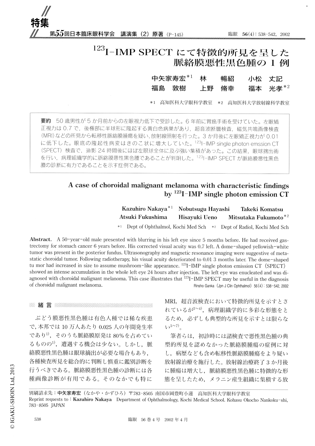
50歳男性が5か月前からの左眼視力低下で受診した。6年前に胃癌手術を受けていた。左眼矯正視力は0.7で,後極部に半球形に隆起する黄白色病巣があり,超音波断層検査,磁気共鳴画像検査(MRI)などの所見から転移性脈絡膜腫瘍を疑い,放射線照射を行った。3か月後に左眼矯正視力が0.01に低下した。眼底の隆起性病変はきのこ状に増大していた。123I-IMP single photon emssion CT(SPECT)検査で,造影24時間後にほぼ左眼球全体に及ぶ強い集積があった。この結果,眼球摘出術を行い,病理組織学的に脈絡膜悪性黒色腫であることが判朋した。123I-IMP SPECTが脈絡膜悪性黒色腫の診断に有力であることを示す症例である。
A 50-year-old male presented with blurring in his left eye since 5 months before. He had received gas-trectomy for stomach cancer 6 years before. His corrected visual acuity was 0.7 left. A dome-shaped yellowish-white tumor was present in the posterior fundus. Ultrasonography and magnetic resonance imaging were suggestive of meta-static choroidal tumor. Following radiotherapy, his visual acuity deteriorated to 0.01 3 months later. The dome-shaped tu mor had increased in size to assume mushroom-like appearance. 123I-IMP single photon emission CT (SPECT) showed an intense accumulation in the whole left eye 24 hours after injection. The left eye was enucleated and was di-agnosed with choroidal malignant melanoma. This case illustrates that 123I-IMP SPECT may be useful in the diagnosis of choroidal malignant melanoma.

Copyright © 2002, Igaku-Shoin Ltd. All rights reserved.


