Japanese
English
- 有料閲覧
- Abstract 文献概要
- 1ページ目 Look Inside
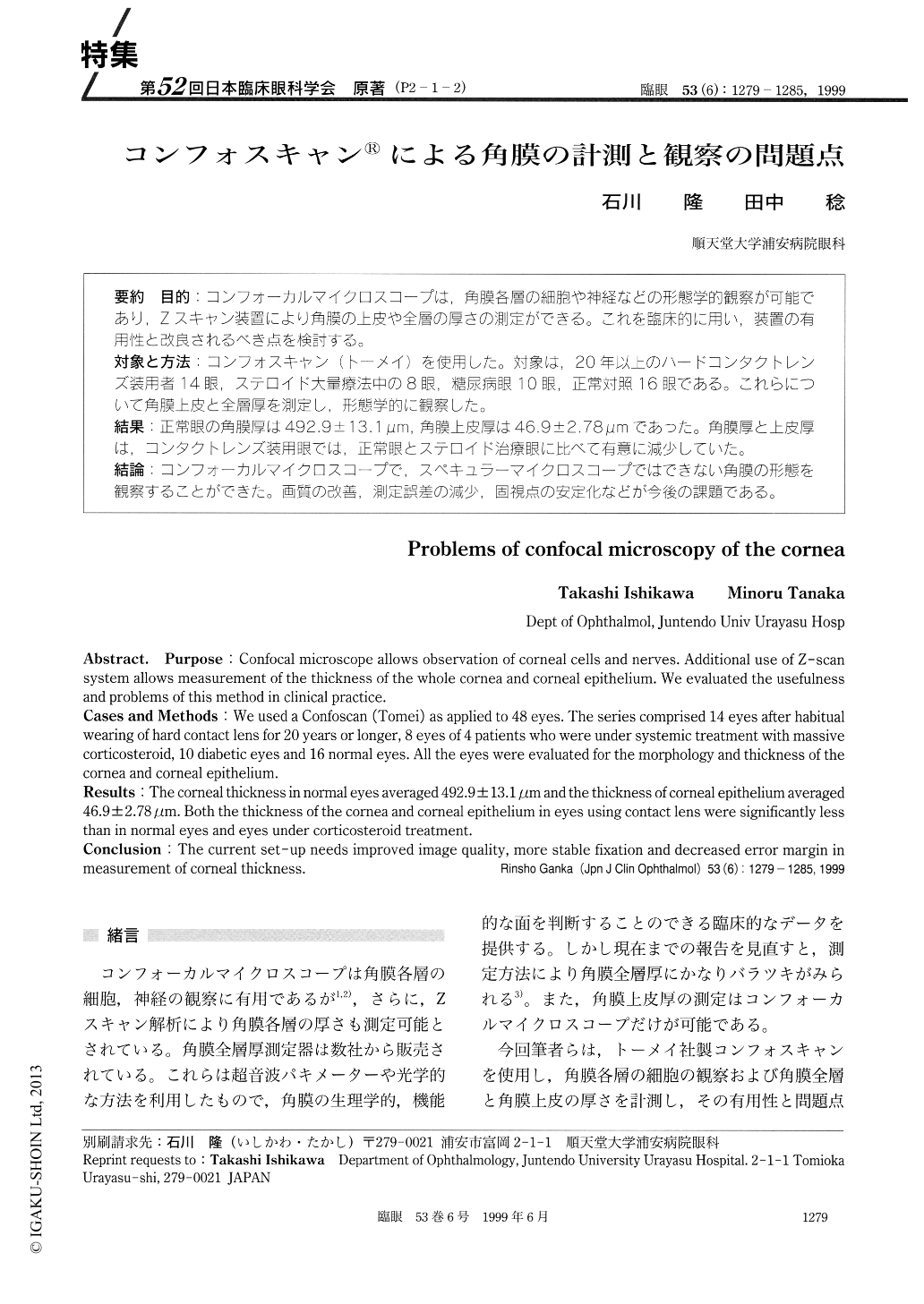
(P2-1-2) 目的:コンフォーカルマイクロスコープは,角膜各層の細胞や神経などの形態学的観察が可能であり,Zスキャン装置により角膜の上皮や全層の厚さの測定ができる。これを臨床的に用い,装置の有用性と改良されるべき点を検討する。
対象と方法:コンフォスキャン(トーメイ)を使用した。対象は,20年以上のハードコンタクトレンズ装用者14眼,ステロイド大量療法中の8眼,糖尿病眼10眼,正常対照16眼である。これらについて角膜上皮と全層厚を測定し,形態学的に観察した。
結果:正常眼の角膜厚は492.9±13.1μm,角膜上皮厚は46.9±2.78μmであった。角膜厚と上皮厚は,コンタクトレンズ装用眼では,正常眼とステロイド治療眼に比べて有意に減少していた。
結論:コンフォーカルマイクロスコープで,スペキュラーマイクロスコープではできない角膜の形態を観察することができた。画質の改善,測定誤差の減少,固視点の安定化などが今後の課題である。
Purpose : Confocal microscope allows observation of corneal cells and nerves. Additional use of Z-scan system allows measurement of the thickness of the whole cornea and corneal epithelium. We evaluated the usefulness and problems of this method in clinical practice.
Cases and Methods : We used a Confoscan (Tomei) as applied to 48 eyes. The series comprised 14 eyes after habitual wearing of hard contact lens for 20 years or longer, 8 eyes of 4 patients who were under systemic treatment with massive corticosteroid, 10 diabetic eyes and 16 normal eyes. All the eyes were evaluated for the morphology and thickness of the cornea and corneal epithelium.
Results : The corneal thickness in normal eyes averaged 492.9±13.1μm and the thickness of corneal epithelium averaged 46.9±2.78μm. Both the thickness of the cornea and corneal epithelium in eyes using contact lens were significantly less than in normal eyes and eyes under corticosteroid treatment.
Conclusion : The current set-up needs improved image quality, more stable fixation and decreased error margin in measurement of corneal thickness.

Copyright © 1999, Igaku-Shoin Ltd. All rights reserved.


