Japanese
English
- 有料閲覧
- Abstract 文献概要
- 1ページ目 Look Inside
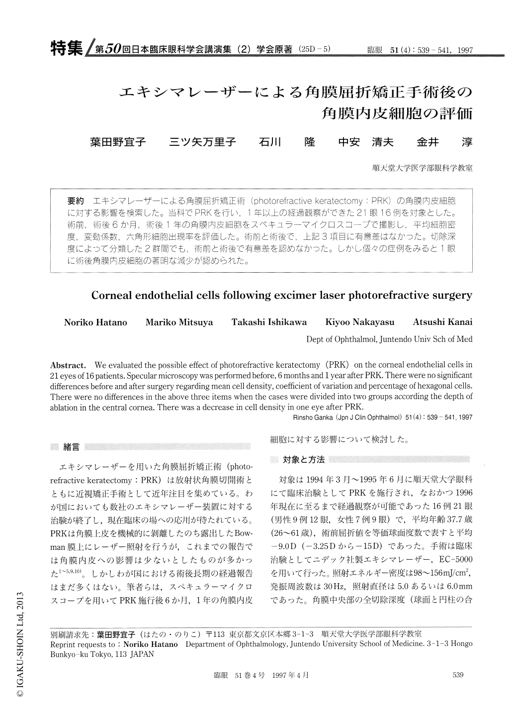
(25D-5) エキシマレーザーによる角膜屈折矯正術(photorefractive keratectomy:PRK)の角膜内皮細胞に対でする影響を検索した。当科でPRKを行い,1年以上の経過観察ができた21眼16例を対象とした。術前,術後6か月,術後1年の角膜内皮細胞をスペキュラーマイクロスコープで撮影し,平均細胞密度,変動係数,六角形細胞出現率を評価した。術前と術後で、上記3項目に有意差はなかった。切除深度によって分類した2群間でも,術前と術後で有意差を認めなかった。しかし個々の症例をみると1眼に術後角膜内皮細胞の著明な減少が認められた。
We evaluated the possible effect of photorefractive keratectomy (PRK) on the corneal endothelial cells in 21 eyes of 16 patients. Specular microscopy was performed before, 6 months and 1 year after PRK. There were no significant differences before and after surgery regarding mean cell density, coefficient of variation and percentage of hexagonal cells.There were no differences in the above three items when the cases were divided into two groups according the depth of ablation in the central cornea. There was a decrease in cell density in one eye after PRK.

Copyright © 1997, Igaku-Shoin Ltd. All rights reserved.


