Japanese
English
- 有料閲覧
- Abstract 文献概要
- 1ページ目 Look Inside
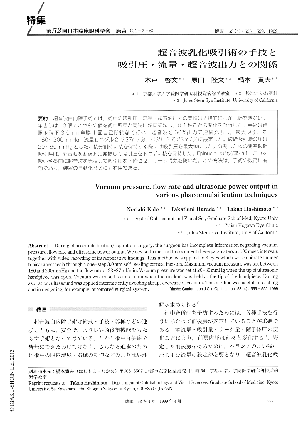
(C1-2-6) 超音波白内障手術では,術中の吸引圧・流量・超音波出力の実情は間接的にしか把握できない。筆者らは,3眼でこれらの値を術中所見と同時に録画記録し,0.1秒ごとの変化を解析した。手術は点眼麻酔下3.Omm角膜1面自己閉鎖創で行い,超音波を60%出力で連続発振し,最大吸引圧を180〜200mmHg,流量をペダル2で27ml/分,ペダル3で23ml/分に設定した。破砕吸引時の圧は20〜80mmHgとした。核分割時に核を保持する際には吸引圧を最大値にした。分割した核の閉塞破砕吸引時は,超音波を断続的に発振して吸引圧を下げずに核を保持した。Epinudeusの処理では,これを吸いきる前に超音波を発振して吸引圧を下降させ,サージ現象を防いだ。この方法は,手術の教育に有効であり,装置の自動化などにも有用である。
During phacoemulsification/aspiration surgery, the surgeon has incomplete information regarding vacuum pressure, flow rate and ultrasonic power output. We devised a method to document these paramaters at 100 msec intervals together with video recording of intraoperative findings. This method was applied to 3 eyes which were operated under topical anesthesia through a one-step 3.0mm self-sealing corneal incision. Maximum vacuum pressure was set between 180 and 200 mmHg and the flow rate at 23-27 ml/min. Vacuum pressure was set at 20-80mmHg when the tip of ultrasonic handpiece was open. Vacuum was raised to maximum when the nucleus was held at the tip of the handpiece. During aspiration, ultrasound was applied intermittently avoiding abrupt decrease of vacuum. This method was useful in teaching and in designing, for example, automated surgical system.

Copyright © 1999, Igaku-Shoin Ltd. All rights reserved.


