Japanese
English
- 有料閲覧
- Abstract 文献概要
- 1ページ目 Look Inside
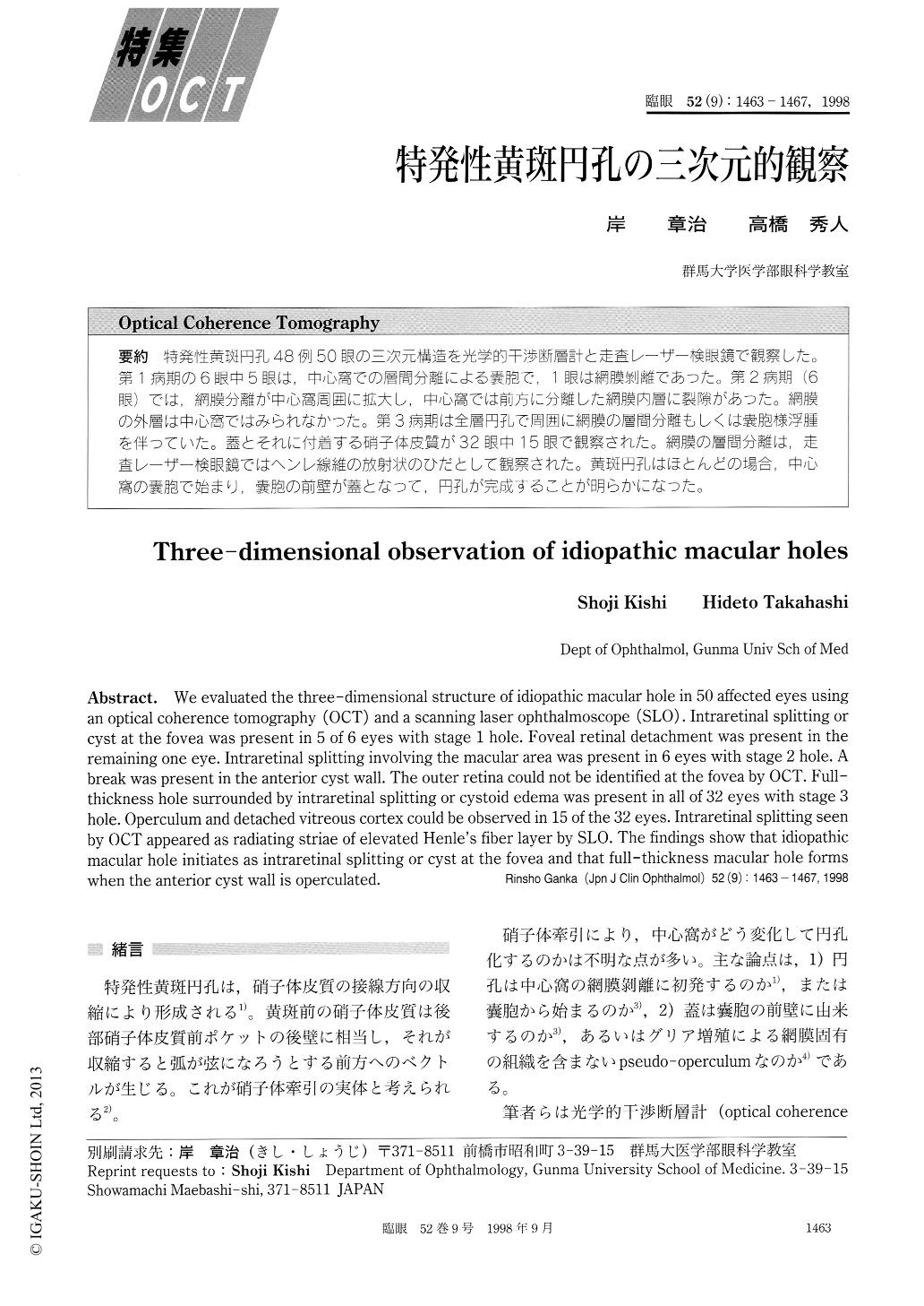
特発性黄斑円孔48例50眼の三次元構造を光学的干渉断層計と走査レーザー検眼鏡で観察した。第1病期の6眼中5眼は,中心窩での層間分離による嚢胞で,1眼は網膜剥離であった。第2病期(6眼)では,網膜分離が中心窩周囲に拡大し,中心窩では前方に分離した網膜内層に裂隙があった。網膜の外層は中心窩ではみられなかった。第3病期は全層円孔で周囲に網膜の層間分離もしくは嚢胞様浮腫を伴っていた。蓋とそれに付着する硝子体皮質が32眼中15眼で観察された。網膜の層間分離は,走査レーザー検銀鏡ではヘンレ線維の放射状のひだとして観察された。黄斑円孔はほとんどの場合,中心窩の嚢胞で始まり,嚢胞の前壁が蓋となって,円孔が完成することが明らかになった。
We evaluated the three-dimensional structure of idiopathic macular hole in 50 affected eyes using an optical coherence tomography (OCT) and a scanning laser ophthalmoscope (SLO) . Intraretinal splitting or cyst at the fovea was present in 5 of 6 eyes with stage 1 hole. Foveal retinal detachment was present in the remaining one eye. Intraretinal splitting involving the macular area was present in 6 eyes with stage 2 hole. A break was present in the anterior cyst wall. The outer retina could not be identified at the fovea by OCT. Full-thickness hole surrounded by intraretinal splitting or cystoid edema was present in all of 32 eyes with stage 3 hole. Operculum and detached vitreous cortex could be observed in 15 of the 32 eyes. Intraretinal splitting seen by OCT appeared as radiating striae of elevated Henle's fiber layer by SLO. The findings show that idiopathic macular hole initiates as intraretinal splitting or cyst at the fovea and that full-thickness macular hole forms when the anterior cyst wall is operculated.

Copyright © 1998, Igaku-Shoin Ltd. All rights reserved.


