Japanese
English
- 有料閲覧
- Abstract 文献概要
- 1ページ目 Look Inside
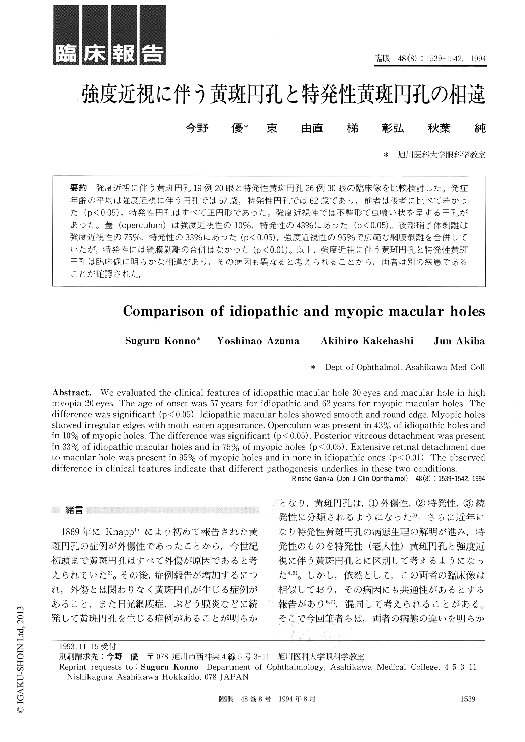
強度近視に伴う黄斑円孔19例20眼と特発性黄斑円孔26例30眼の臨床像を比較検討した。発症年齢の平均は強度近視に伴う円孔では57歳,特発性円孔では62歳であり,前者は後者に比べて若かった(p<0.05)。特発性円孔はすべて正円形であった。強度近視性では不整形で虫喰い状を呈する円孔があった。蓋(operculum)は強度近視性の10%,特発性の43%にあった(p<0.05)。後部硝子体剥離は強度近視性の75%,特発性の33%にあった(p<0.05)。強度近視性の95%で広範な網膜剥離を合併していたが,特発性には網膜剥離の合併はなかった(p<0.01)。以上,強度近視に伴う黄斑円孔と特発性黄斑円孔は臨床像に明らかな相違があり,その病因も異なると考えられることから,両者は別の疾患であることが確認された。
We evaluated the clinical features of idiopathic macular hole 30 eyes and macular hole in high myopia 20 eyes. The age of onset was 57 years for idiopathic and 62 years for myopic macular holes. The difference was significant (p<0.05). Idiopathic macular holes showed smooth and round edge. Myopic holes showed irregular edges with moth-eaten appearance. Operculum was present in 43% of idiopathic holes and in 10% of myopic holes. The difference was significant (p<0.05). Posterior vitreous detachment was present in 33% of idiopathic macular holes and in 75% of myopic holes (p<0.05). Extensive retinal detachment due to macular hole was present in 95% of myopic holes and in none in idiopathic ones (p <0.01). The observed difference in clinical features indicate that different pathogenesis underlies in these two conditions.

Copyright © 1994, Igaku-Shoin Ltd. All rights reserved.


