Japanese
English
- 有料閲覧
- Abstract 文献概要
- 1ページ目 Look Inside
乱視矯正角膜切開術(asitigmatic kerato—tomy,以下AK)後早期の角膜形状変化を角膜形状解析装置(TMS-1:Computed Anatomy社)により検討した。
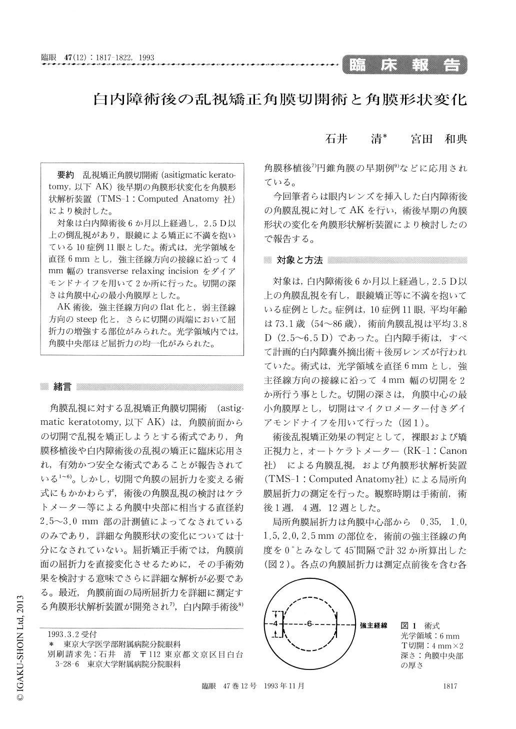
対象は白内障術後6か月以上経過し,2.5D以上の倒乱視があり,眼鏡による矯正に不満を抱いている10症例11眼とした。術式は,光学領域を直径6mmとし,強主径線方向の接線に沿って4mm幅のtransverse relaxing incisionをダイアモンドナイフを用いて2か所に行った。切開の深さは角膜中心の最小角膜厚とした。
AK術後,強主径線方向のflat化と,弱主径線方向のsteep化と,さらに切開の両端において屈折力の増強する部位がみられた。光学領域内では,角膜中央部ほど屈折力の均一化がみられた。
We evaluated the changes in corneal topography following astigmatic keratotomy in 11 pseudo-phakic eyes. We used automated keratometer by Canon Inc. and computerized corneal shape analyzer by Computed Anatomy Inc. All the eyes had undergone posterior chamber implantation and had astigmatism-against-the-rule by 2.5 diopters (D) or greater at 6 months or longer after surgery. Two parallel 4-mm transverse relaxing incisions were placed perpendicular to the steep meridian 3 mm from the center of the cornea. At 12 weeks after astigmatic keratotomy, the astigmatism chan-ged from the preoperative average of 3.8D to 2.0D. The central corneal area became flatter.

Copyright © 1993, Igaku-Shoin Ltd. All rights reserved.


