Japanese
English
- 有料閲覧
- Abstract 文献概要
- 1ページ目 Look Inside
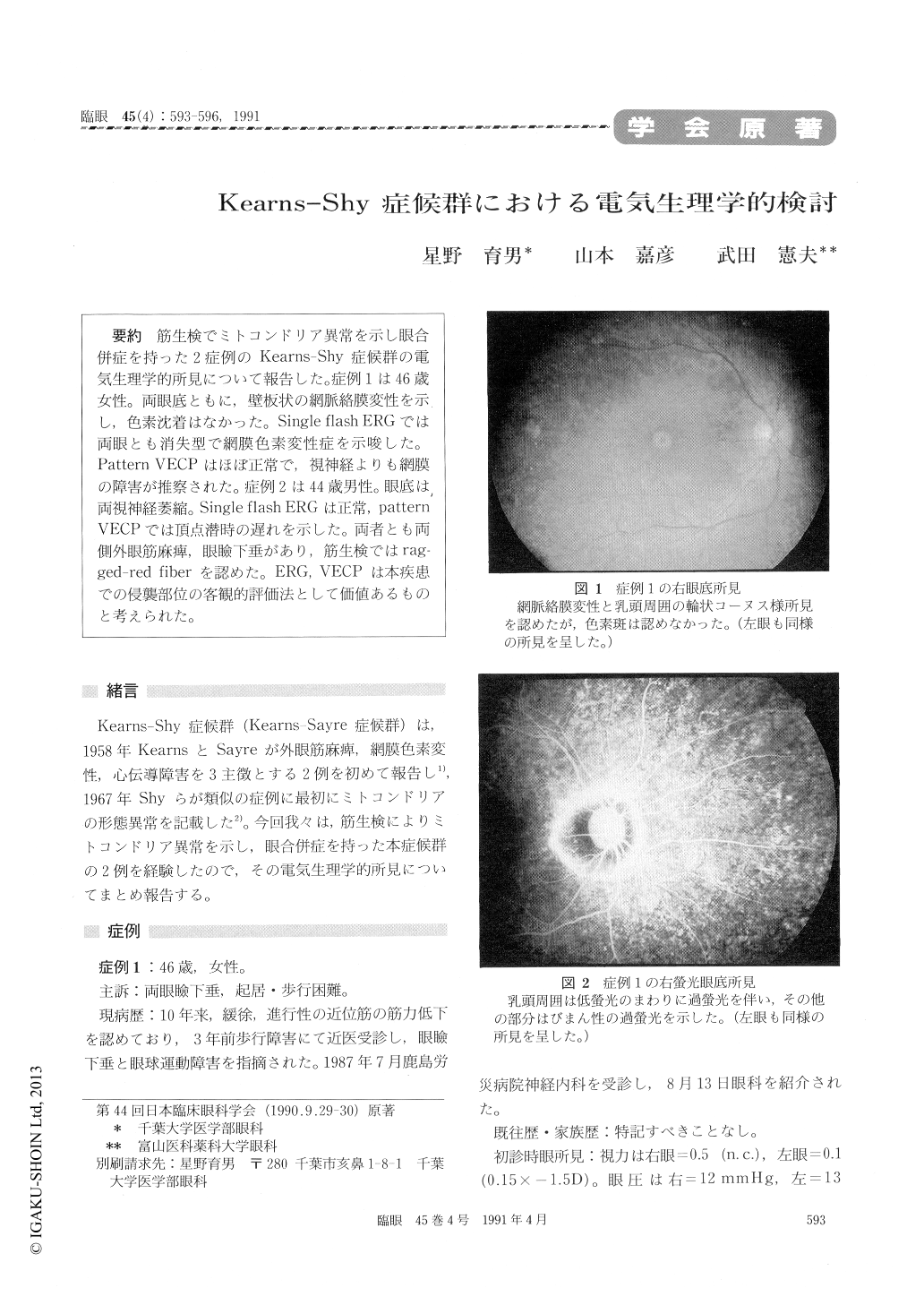
筋生検でミトコンドリア異常を示し眼合併症を持った2症例のKearns-Shy症候群の電気生理学的所見について報告した。症例1は46歳女性。両眼底ともに,壁板状の網脈絡膜変性を示し,色素沈着はなかった。Single flash ERGでは両眼とも消失型で網膜色素変性症を示唆した。Pattern VECPはほぼ正常で,視神経よりも網膜の障害が推察された。症例2は44歳男性。眼底は,両視神経萎縮。Single flash ERGは正常,pattern VECPでは頂点潜時の遅れを示した。両者とも両側外眼筋麻痺,眼瞼下垂があり,筋生検ではrag-ged-red fiberを認めた。ERG,VECPは本疾患での侵襲部位の客観的評価法として価値あるものと考えられた。
We studied electrophysiologically on two cases of the Kearns-Shy syndrome, in which some struc-tural abnormalities of mitochondria were shown.
A 46-year-old female presented with bilateral ptosis, total external ophthalmoplegia, neurosen-sory hearing loss and muscular weakness. She manifested the fundus feature of tapetoretinal degeneration and no hyper pigmentation. The sin-gle-flash electroretinograms (ERGs) were almost extinguished. The pattern visually evoked cortical potentials (PVECPs) were almost normal.
A 46-year-old male presented with bilateral ptosis, total external ophthalmoplegia, neurosen-sory hearing loss and elevated CSF protein level. Both fundi showed optic atrophy. The single-flash ERGs were almost normal. The peak latency of the P100 of the PVECP was delayed.
In both cases, “ragged red fibers” were observed in muscle biopsy specimens with modified Gomori trichrome stain.
Thus the electrophysiological studies may pro-vide us a way of evaluating the damaged system of visual function in the Kearns-Shy syndrome.

Copyright © 1991, Igaku-Shoin Ltd. All rights reserved.


