Japanese
English
- 有料閲覧
- Abstract 文献概要
- 1ページ目 Look Inside
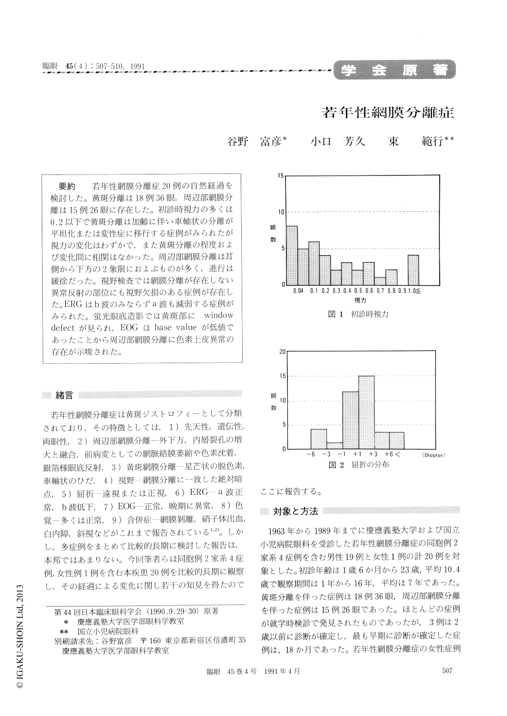
若年性網膜分離症20例の自然経過を検討した。黄斑分離は18例36眼,周辺部網膜分離は15例26眼に存在した。初診時視力の多くは0.2以下で黄斑分離は加齢に伴い車軸状の分離が平坦化または変性症に移行する症例がみられたが視力の変化はわずかで,また黄斑分離の程度および変化間に相関はなかった。周辺部網膜分離は耳側から下方の2象限におよぶものが多く,進行は緩徐だった。視野検査では網膜分離が存在しない異常反射の部位にも視野欠損のある症例が存在した。ERGはb波のみならずa波も減弱する症例がみられた。蛍光眼底造影では黄斑部にwindow defectが見られ,EOGはbase valueが低値であったことから周辺部網膜分離に色素上皮異常の存在が示唆された。
We reviewed the natural course of 20 cases of X-linked juvenile retinoschisis. Foveal retinoschisis was present in 36 eyes, 18 cases. Peripheral retinos-chisis was present in 26 eyes, 15 cases. The visual acuity was less than 0.2 in the majority of cases.
Initial radiating folds in foveal retinoschisis became flatter or turned into cystic degeneration later. The visual acuity was not independent of the degree or clinical pattern of foveal retinoschisis.
Peripheral retinoschisis was of slow progression and was frequenty located in the inferotemporal quadrant. Visual field defect corresponded not only to the area of peripheral retinoschisis but also to that of abnormal silver-grey reflex of the retina.
Electroretinogram showed low voltage in base value and decrease in b wave and occasionally in a wave. Fluorescein angiography showed window defects in the macula and abnormalities in the pigment epithelium in areas of peripheral retinos-chisis.

Copyright © 1991, Igaku-Shoin Ltd. All rights reserved.


