Japanese
English
- 有料閲覧
- Abstract 文献概要
- 1ページ目 Look Inside
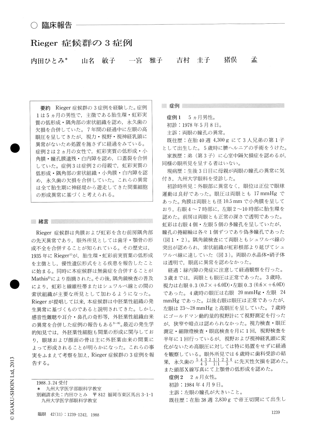
Rieger症候群の3症例を経験した.症例1は5カ月の男性で,主徴である胎生環・虹彩実質の低形成・隅角部の索状組織を認め,永久歯の欠損を合併していた.7年間の経過中に左眼の高眼圧を呈してきたが,視力・視野・視神経乳頭に異常がないため処置を施さずに経過をみている.症例2は2カ月の女性で,虹彩実質の低形成・小角膜・瞳孔膜遺残・白内障を認め,口蓋裂を合併していた.症例3は症例2の母親で,虹彩実質の低形成・隅角部の索状組織・小角膜・白内障を認め,永久歯の欠損を合併していた.これらの異常は全て胎生期に神経堤から遊走してきた間葉細胞の形成異常に基づくと考えられる.
We observed Rieger's syndrome in 3 patients : a 5 -month-old male, 2-month-old female and 24-year -old female. The first case manifested posterior embryotoxon, hypoplasia of iris, and strands bridg-ing the iridocorneal angle, as well as polycoria, microcornea and congenital absence of tooth. Ocu-lar hypertension developed at the age of 4 years, without particular changes in visual acuity or visual field during the following 3 years. The sec-ond case manifested hypoplasia of iris, mi-crocornea, persistent pupillary membrane, cataract, strabismus and dysgnathia. Posterior embryotoxon was absent. The third case, her mother, manifested hypoplasia of iris, iris strands bridging iridocorneal angle, microcornea, cataract, glaucoma and absence of tooth. Posterior embryotoxon was absent.
All the three cases manifested, as common fea-tures of the disease, goniodysgenesis involving the cornea and iris, absence of tooth or dysgnathia. These abnormalities seemed to be clinical manifes-tations of dysplasia of mesenchymal tissue derived from the neural crest.
Rinsho Ganka (Jpn J Clio Ophthalmol) 42(11) : 1239-1242, 1988

Copyright © 1988, Igaku-Shoin Ltd. All rights reserved.


