Japanese
English
- 有料閲覧
- Abstract 文献概要
- 1ページ目 Look Inside
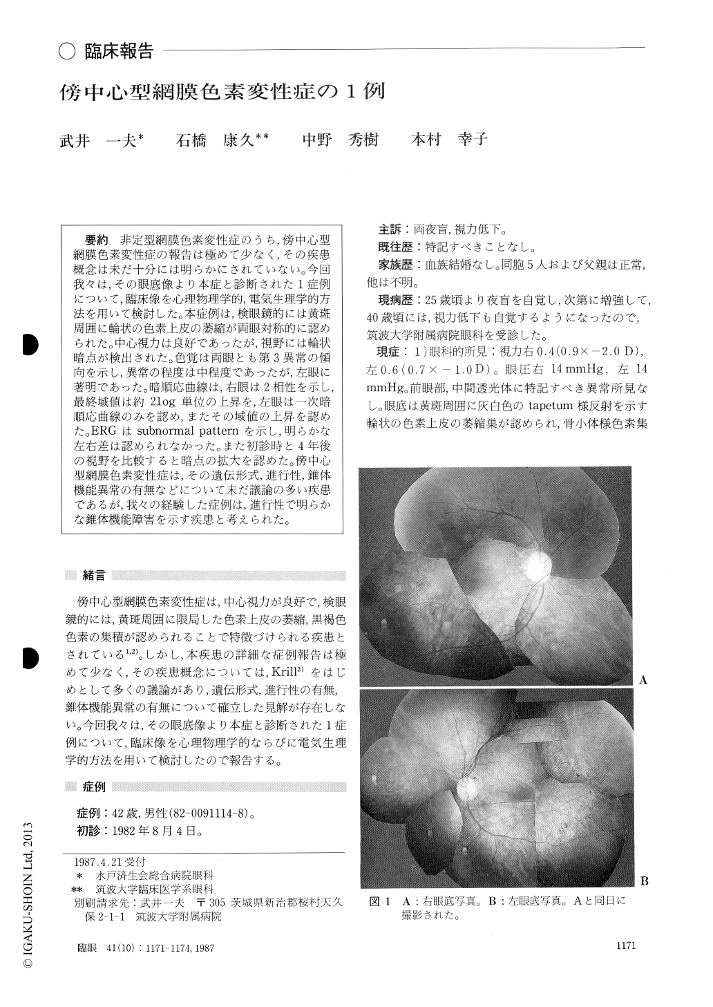
非定型網膜色素変性症のうち,傍中心型網膜色素変性症の報告は極めて少なく,その疾患概念は未だ十分には明らかにされていない.今回我々は,その眼底像より本症と診断された1症例について,臨床像を心理物理学的,電気生理学的方法を用いて検討した.本症例は,検眼鏡的には黄斑周囲に輪状の色素上皮の萎縮が両眼対称的に認められた.中心視力は良好であったが,視野には輪状暗点が検出された.色覚は両眼とも第3異常の傾向を示し,異常の程度は中程度であったが,左眼に著明であった.暗順応曲線は,右眼は2相性を示し,最終域値は約2log単位の上昇を,左眼は一次暗順応曲線のみを認め,またその域値の上昇を認めた.ERGはsubnormal patternを示し,明らかな左右差は認められなかった.また初診時と4年後の視野を比較すると暗点の拡大を認めた.傍中心型網膜色素変性症は,その遺伝形式,進行性,錐体機能異常の有無などについて未だ議論の多い疾患であるが,我々の経験した症例は,進行性で明らかな錐体機能障害を示す疾患と考えられた.
A 42-year-old male was diagnosed as pericentral pigmentary retinal degeneration in both eyes. Night blindness had been present since the age of 25 years. The fundus was characterized by annular depig-mented zone around the macula. The visual acuity was well retained during the 4-year follow-up period. The visual fields showed paracentralscotoma. Color vision tests showed moderate tritan defect. Dark adaptation and electroretinogram were abnormal and suggestive of dysfunction in the rod the cone system. The pericentral lesion extended slowly towards the periphery and the fovea during the 4 years. The mode of inheritance was inconclusive. The case seemed to represent a rare atypical form of pigmentary retinal degenera-tion.
Rinsho Ganka (Jpn J Clin Ophthalmol) 41(10) : 1171-1174, 1987

Copyright © 1987, Igaku-Shoin Ltd. All rights reserved.


