Japanese
English
- 有料閲覧
- Abstract 文献概要
- 1ページ目 Look Inside
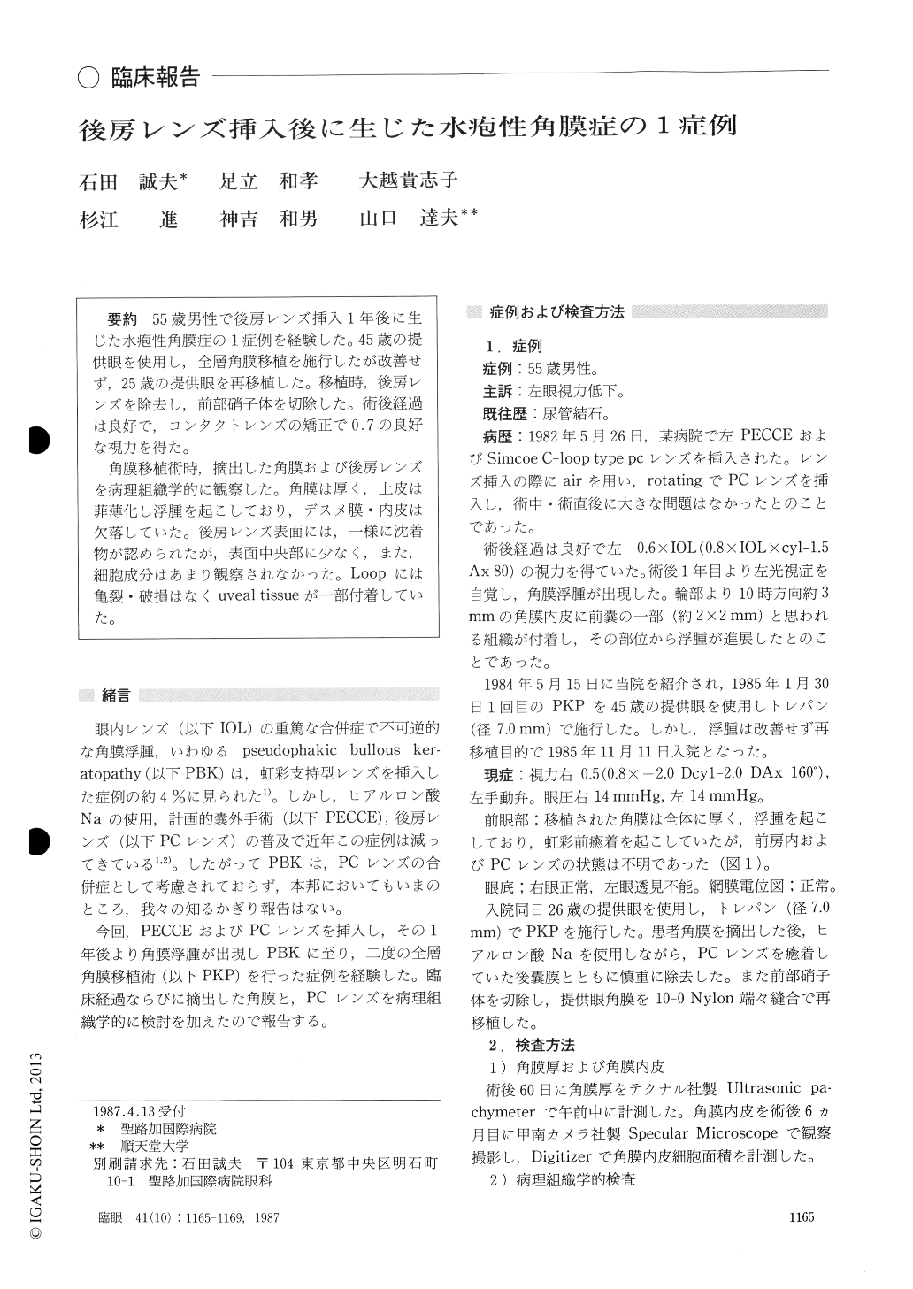
55歳男性で後房レンズ挿入1年後に生じた水疱性角膜症の1症例を経験した.45歳の提供眼を使用し,全層角膜移植を施行したが改善せず,25歳の提供眼を再移植した.移植時,後房レンズを除去し,前部硝子体を切除した.術後経過は良好で,コンタクトレンズの矯正で0.7の良好な視力を得た.
角膜移植術時,摘出した角膜および後房レンズを病理組織学的に観察した.角膜は厚く,上皮は菲薄化し浮腫を起こしており,デスメ膜・内皮は欠落していた.後房レンズ表面には,一様に沈着物が認められたが,表面中央部に少なく,また,細胞成分はあまり観察されなかった.Loopには亀裂・破損はなくuveal tissueが一部付着していた.
A 55-year-old male developed bullous ker-atopathy one year after extracapsular cataract extraction with implantation of Simcoe C-loop posterior chamber intraocular lens in his left eye. The implanted lens was not in touch with the posterior corneal surface. Penetrating keratoplasty without removal of the intraocular lens failed because of graft opacity. A second keratoplasty was performed, 3 and a half years after the initial cataract extraction, with removal of the intraocular lens and anterior vitrectomy. Thedonor cornea was obtained from a 26-year-old person. The graft stayed clear with the visual acuity of 0.7 corrected with hard contact lens. We observed the removed cornea and the intraocular lens by light and scanning electron microscopy. The corneal button was thick with prominent intercellular spaces of epithelial cells. The epithelial layer was thin. Endothelial cells were absent. The intraocular lens did not manifest gross abnormalities as macrophage-like cells on its surface, peeling, cracks or biodegraded changes except uveal tissue attached to the haptics. The cause of the pseudophakic bullous keratopathy could not be determined.
Rinsho Ganka (Jpn J Clin Ophthalmol) 41(10) : 1165-1169, 1987

Copyright © 1987, Igaku-Shoin Ltd. All rights reserved.


