Japanese
English
- 有料閲覧
- Abstract 文献概要
- 1ページ目 Look Inside
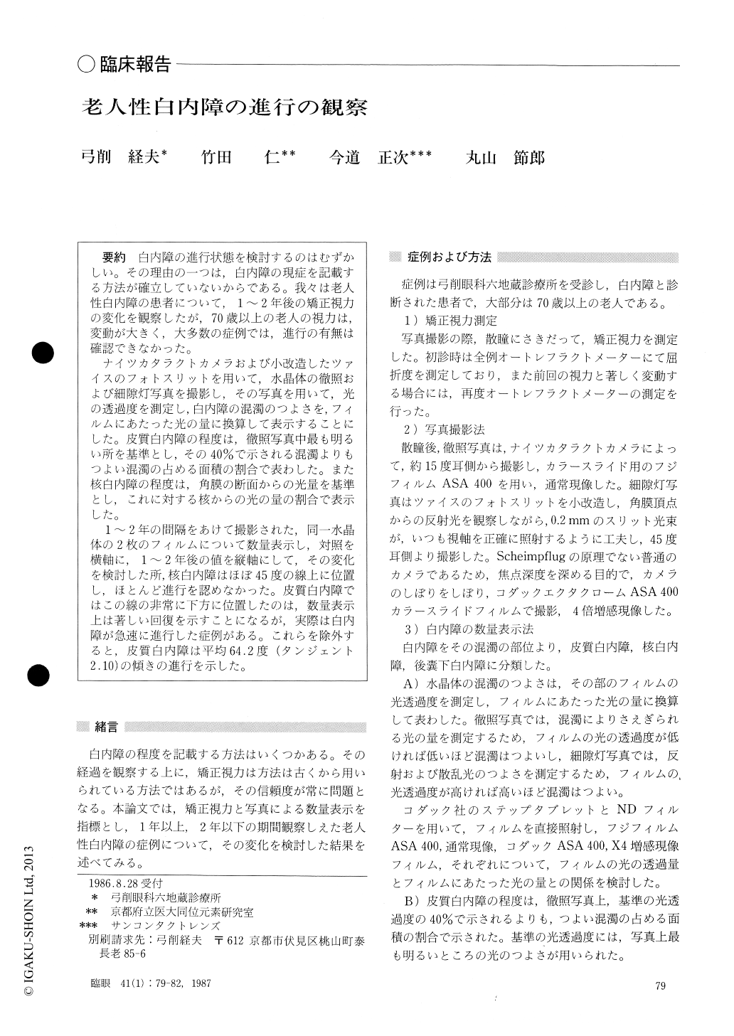
白内障の進行状態を検討するのはむずかしい.その理由の一つは,白内障の現症を記載する方法が確立していないからである.我々は老人性白内障の患者について,1〜2年後の矯正視力の変化を観察したが,70歳以上の老人の視力は,変動が大きく,大多数の症例では,進行の有無は確認できなかった.
ナイツカタラクトカメラおよび小改造したツァイスのフォトスリットを用いて,水晶体の徹照および細隙灯写真を撮影し,その写真を用いて,光の透過度を測定し,白内障の混濁のつよさを,フィルムにあたった光の量に換算して表示することにした.皮質白内障の程度は,徹照写真中最も明るい所を基準とし,その40%で示される混濁よりもつよい混濁の占める面積の割合で表わした.また核白内障の程度は,角膜の断面からの光量を基準とし,これに対する核からの光の量の割合で表示した.
1〜2年の間隔をあけて撮影された,同一水晶体の2枚のフィルムについて数量表示し,対照を横軸に,1〜2年後の値を縦軸にして,その変化を検討した所,核白内障はほぼ45度の線上に位置し,ほとんど進行を認めなかった.皮質白内障ではこの線の非常に下方に位置したのは,数量表示上は著しい回復を示すことになるが,実際は白内障が急速に進行した症例がある.これらを除外すると,皮質白内障は平均64.2度(タンジェント2.10)の傾きの進行を示した.
In order to objectively evaluate the status of senile cataract, we photographed the crystalline lens by retroillumination and slitlamp image, the former with a Neitz cataract camera and the latter with a Zeiss photoslitlamp. The intensity of lens opacity was expres-sed as the amount of light reaching the photographed film. The severity of cortical cataract was expressed as percentage of the area occupied by opaque regions greater than 40 % of the background illumination in the retroilluminated photograph. In nuclear cataract, the severity was expressed as the amount of scattered light from the nucleus with respect to the corneal section in the slitlamp photograph.
We applied this method to eyes with senile cataract. When the same eyes were examined after an interval of 1 to 2 years by the method, a significant progression was noted in eyes with cortical cataract. Eyes with senile nuclear cataract remained virtually stationary in severity.
Because of its objective and quantitative nature, our method promises to be of value as a research tool in elucidating the natural history, etiological and thera-peutic aspects of senile cataract.
Rinsho Ganka (Jpn J Clin Ophthalmol) 41(1) : 79-82, 1987

Copyright © 1987, Igaku-Shoin Ltd. All rights reserved.


