Japanese
English
- 有料閲覧
- Abstract 文献概要
- 1ページ目 Look Inside
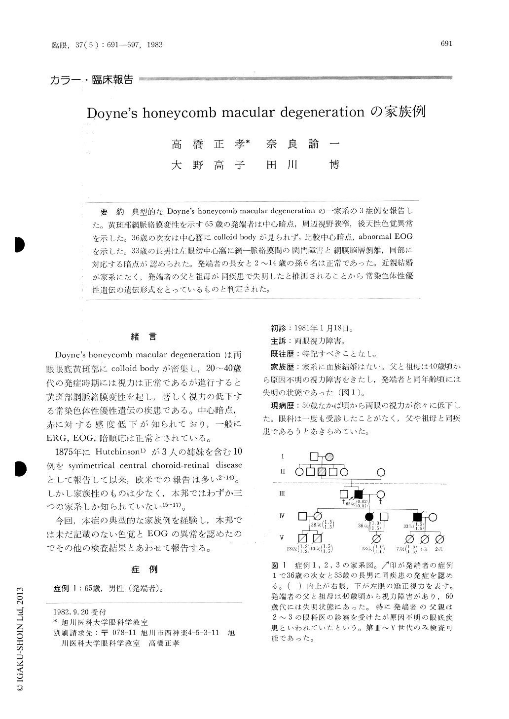
典型的なDoyne's honeycomb macular degenerationの一家系の3症例を報告した。黄斑部網脈絡膜変性を示す65歳の発端者は中心暗点,周辺視野狭窄,後天性色覚異常を示した。36歳の次女は中心窩にcolloid bodyが見られず,比較中心暗点,abnormal EOGを示した。33歳の長男は左眼傍中心窩に網—脈絡膜間の関門障害と網膜脳層剥離,同部に対応する暗点が認められた。発端者の長女と2〜14歳の孫6名は正常であった。近親結婚が家系になく,発端者の父と祖母が同疾患で失明したと推測されることから常染色体性優性遺伝の遺伝形式をとっているものと判定された。
Three members of a family who presented Doyne's honeycomb macular degeneration were clinically studied. A 65-year-old male whose vision had been deteriorating since the fourth decade of his life showed colloid bodies closely grouped in the papillomacular area. These bodies coalesced in the parafoveal region and were replaced by retino-choroidal atrophy with blackish pigmentary patches at the fovea in both eyes.
One of his three children, a 38-year-old daughter, was normal. His 36-year-old daughter with normal vision revealed closely grouped colloid bodies in the papillomacular area sparing the fovea.

Copyright © 1983, Igaku-Shoin Ltd. All rights reserved.


