Japanese
English
- 有料閲覧
- Abstract 文献概要
- 1ページ目 Look Inside
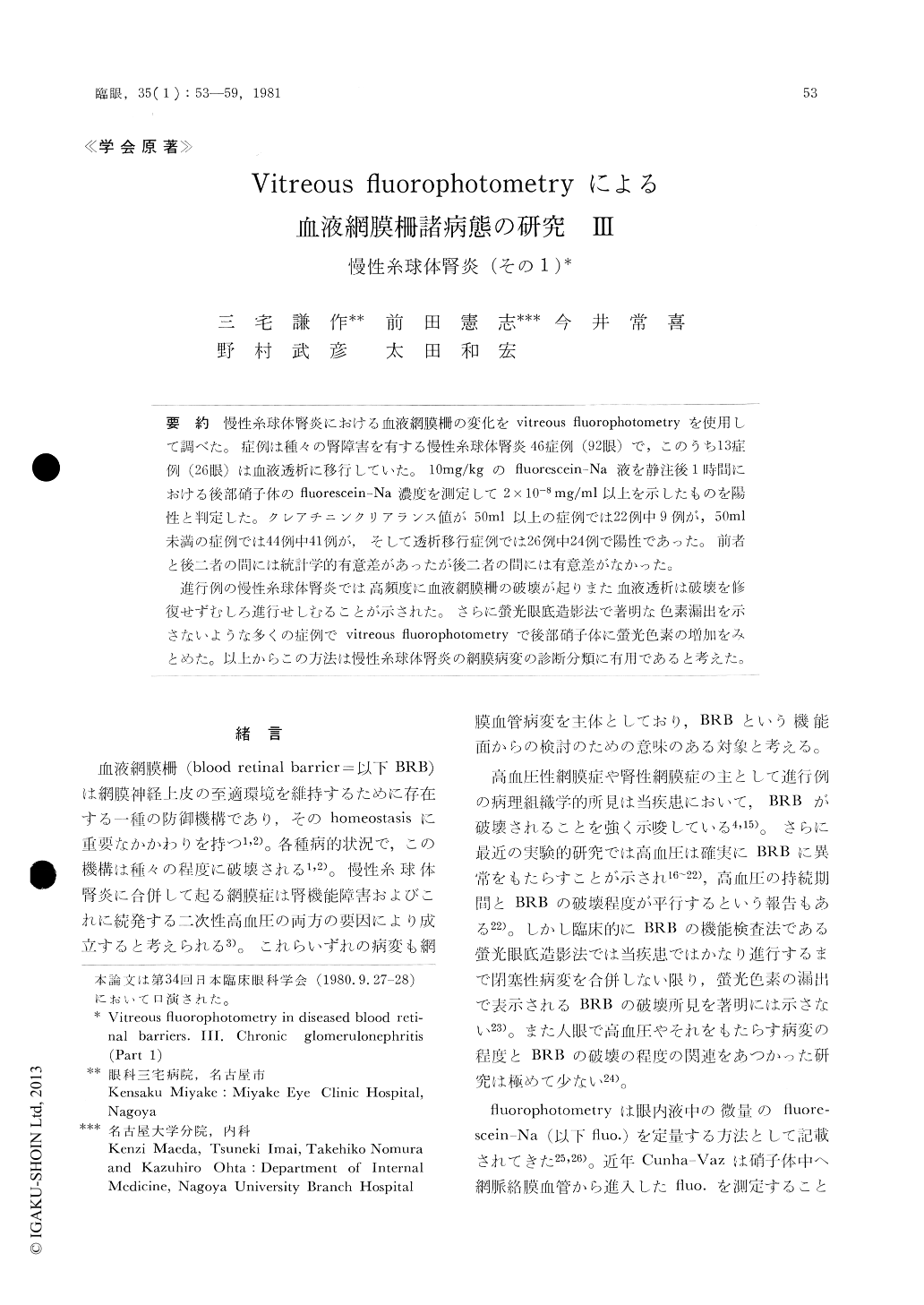
慢性糸球体腎炎における血液網膜柵の変化をvitreous fluorophotometryを使用して調べた。症例は種々の腎障害を有する慢性糸球体腎炎46症例(92眼)で,このうち13症例(26眼)は血液透析に移行していた。10mg/kgのfluorescein-Na液を静注後1時間における後部硝子体のfluorescein-Na濃度を測定して2×10−8mg/ml以上を示したものを陽性と判定した。クレアチニンクリアランス値が50ml以上の症例では22例中9例が,50ml未満の症例では44例中41例が,そして透析移行症例では26例中24例で陽性であった。前者と後二者の間には統計学的有意差があったが後二者の間には有意差がなかった。
進行例の慢性糸球体腎炎では高頻度に血液網膜柵の破壊が起りまた血液透析は破壊を修復せずむしろ進行せしむることが示された。さらに螢光眼底造影法で著明な色素漏出を示さないような多くの症例でvitreous fluorophotometryで後部硝子体に螢光色素の増加をみとめた。以上からこの方法は慢性糸球体腎炎の網膜病変の診断分類に有用であると考えた。
Vitreous fluorophotometry was used to evaluate changes in the blood-retinal barrier to fluorescein sodium in patients with chronic gromeluronephritis. Examined were 46 cases (92 eyes) with various degrees of renal dysfunction 13 cases (26 eyes) were treated by hemodialysis. One hour after intravenous injection of fluorescein sodium, 10mg/ kg, the concentration in the posterior vitreous was measured and the value of 2 × 10⁻⁸mg/ml or more was judged as positive.

Copyright © 1981, Igaku-Shoin Ltd. All rights reserved.


