Japanese
English
- 有料閲覧
- Abstract 文献概要
- 1ページ目 Look Inside
緒言
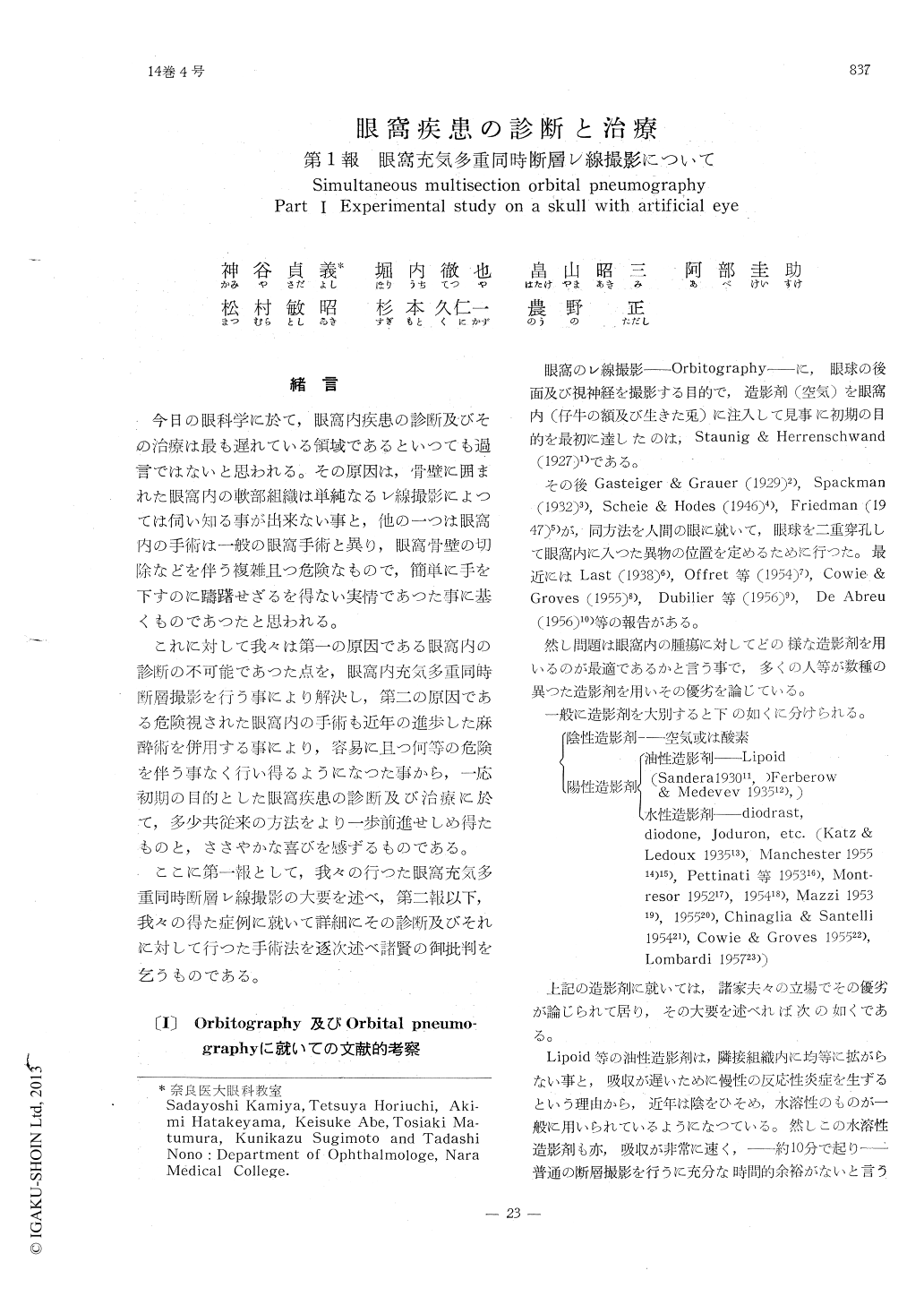
今日の眼科学に於て,眼窩内疾患の診断及びその治療は最も遅れている領域であるといつても過言ではないと思われる。その原因は,骨壁に囲まれた眼窩内の軟部組織は単純なるレ線撮影によつては伺い知る事が出来ない事と,他の一つは眼窩内の手術は一般の眼窩手術と異り,眼窩骨壁の切除などを伴う複雑且つ危険なもので,簡単に手を下すのに躊躇せざるを得ない実情であつた事に基くものであつたと思われる。
これに対して我々は第一の原因である眼窩内の診断の不可能であつた点を,眼窩内充気多重同時断層撮影を行う事により解決し,第二の原因である危険視された眼窩内の手術も近年の進歩した麻酔術を併用する事により,容易に且つ何等の危険を伴う事なく行い得るようになつた事から,一応初期の目的とした眼窩疾患の診断及び治療に於て,多少共従来の方法をより一歩前進せしめ得たものと,ささやかな喜びを感ずるものである。
An experimental study of simultaneous multisection orbital pneumography was performed on a skull with artificial eye, which was filled with water in the ping-pong ball.
The technical factores were 75KV 30mA at 0.5secound. Degrees of exposure were 20, 40 and 60, and the positions of the body to the line of the exposure were 180°, 90° and 45°. The orbital pneumography was taken in the optical foramen position and lateral position, with two exposure of 4 or 5 body-section radiography which was produced simultaneously.

Copyright © 1960, Igaku-Shoin Ltd. All rights reserved.


