Japanese
English
- 有料閲覧
- Abstract 文献概要
- 1ページ目 Look Inside
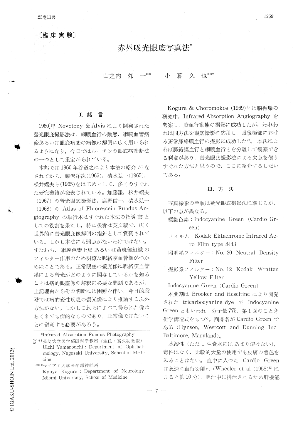
I.緒言
1960年Novotony & Alvisにより開発された螢光眼底撮影法は,網膜血行の動態,網膜血管病変あるいは眼底病変の病像の解明に広く用いられるようになり,今日ではルーチンの眼底病診断法の一つとして重宝がられている。
本邦では1960年谷道之により本法の紹介がなされてから,藤沢洋次(1965),清水弘一(1965),松井端夫ら(1965)をはじめとして,多くのすぐれた研究業績が発表されている。加藤謙,松井端夫(1967)の螢光眼底撮影法,鹿野信一,清水弘一(1968)のAtlas of Fluorescein Fundus An—giographyの単行本はすぐれた本法の指導書としての役割を果たし,特に後者は英文版で,広く世界的に螢光眼底像解明の指針として賞賛されている。しかし本法にも弱点がないわけではない。すなわち,網膜色素上皮あるいは黄斑部組織のフィルター作用のため明瞭な脈絡膜血管像がつかめぬことである。正常眼底の螢光像に脈絡膜血管系による螢光がどのように関与しているかを知ることは病的眼底像の解釈に必要な問題であるが,上記理由からその判断には困難を伴い,今日的段階では病的変性疾患の螢光像により推論する以外方法がない。しかしこれらによつて得られた像はあくまでも病的なものであり,正常像ではないことに留意する必要があろう。
Angiography of the choroidal vessels in owl monkey was conducted with the use of indocy-anine green as contrast media and infrared color film (Kodak Ektachrome Infrared Aero Film type 8443). The dye was rapidly injected into the common carotid through a cannule followed by a rapid serial photography of the fundus filtered by No. 20 neutral density filter and No. 12 Kodak wratten yellow filter.
The dye-appeared in the choroidal vessels two-thirds to one second following injection, initially assuming a "geographical" pattern. The optic disc was stained with the dye beginning at the disc margin.

Copyright © 1969, Igaku-Shoin Ltd. All rights reserved.


