Japanese
English
- 有料閲覧
- Abstract 文献概要
- 1ページ目 Look Inside
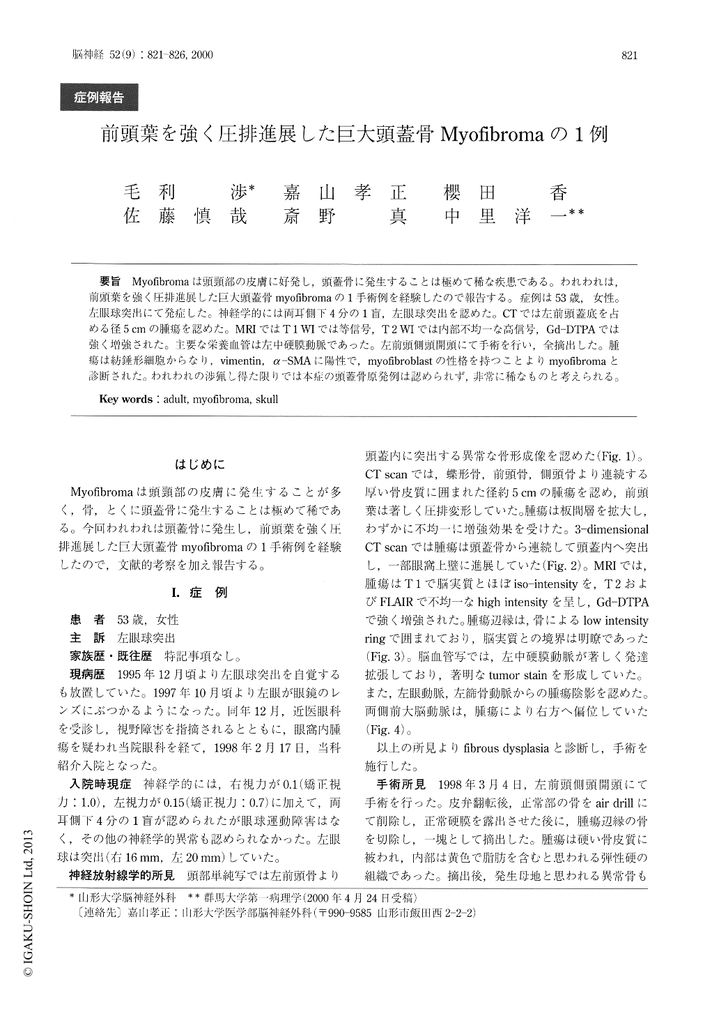
Myofibromaは頭頸部の皮膚に好発し,頭蓋骨に発生することは極めて稀な疾患である。われわれは,前頭葉を強く圧排進展した巨大頭蓋骨myofibromaの1手術例を経験したので報告する。症例は53歳,女性。左眼球突出にて発症した。神経学的には両耳側下4分の1盲,左眼球突出を認めた。CTでは左前頭蓋底を占める径5cmの腫瘍を認めた。MRIではT1WIでは等信号,T2WIでは内部不均一な高信号,Gd-DTPAでは強く増強された。主要な栄養血管は左中硬膜動脈であった。左前頭側頭開頭にて手術を行い,全摘出した。腫瘍は紡錘形細胞からなり,vimentin,α-SMAに陽性で,myofibroblastの性格を持つことよりmyofibromaと診断された。われわれの渉猟し得た限りでは本症の頭蓋骨原発例は認められず,非常に稀なものと考えられる。
We report a rare case of giant skull myofibroma oc-cupying left anterior cranial fossa. A 53-year- old woman presented with left exophthalmos for 2 years. Neurological examination showed left exophthalmos, disturbance of bilateral visual acuity, and bitemporal hemianopsia. A CT scan revealed an ossifing mass at left anterior cranial fossa. On magnetic resonance im-ages, the tumor showed iso-intensity on T 1-weighted image, heterogeneous high intensity on T 2-weighted image, and was heterogeneously well-enhanced afteradministration of Gd-DTPA. The tumor was fed mainly by middle meningeal artery.

Copyright © 2000, Igaku-Shoin Ltd. All rights reserved.


