Japanese
English
- 有料閲覧
- Abstract 文献概要
- 1ページ目 Look Inside
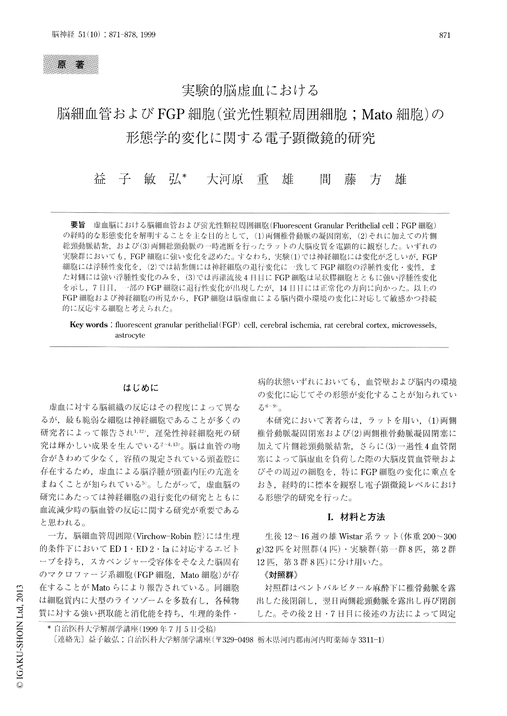
虚血脳における脳細血管および蛍光性顆粒周囲細胞(Fluorescent Granular Perithelial cell;FGP細胞)の経時的な形態変化を解明することを主な目的として,(1)両側椎骨動脈の凝固閉塞,(2)それに加えての片側総頸動脈結紮,および(3)両側総頸動脈の一時遮断を行ったラットの大脳皮質を電顕的に観察した。いずれの実験群においても,FGP細胞に強い変化を認めた。すなわち,実験(1)では神経細胞には変化が乏しいが,FGP細胞には浮腫性変化を,(2)では結紮側には神経細胞の退行変化に一致してFGP細胞の浮腫性変化・変性,また対側には強い浮腫性変化のみを,(3)では再灌流後4日目にFGP細胞は星状膠細胞とともに強い浮腫性変化を示し,7日目,一部のFGP細胞に退行性変化が出現したが,14日目には正常化の方向に向かった。以上のFGP細胞および神経細胞の所見から,FGP細胞は脳虚血による脳内微小環境の変化に対応して敏感かつ持続的に反応する細胞と考えられた。
In order to clarify the sequential changes of the morphology of vascular cells and FGP cells under cerebral ischemia, 32 male Wistar rats were em-ployed. The FGP cells in the present paper are distrib-uted along cerebral microvessels, and markedly potent in the uptake capacity for endo-and exogenous sub-stances under the physiological and pathological con-ditions.
Under the anesthesia of pentobarbital, experimental animals suffered from cerebral ischemia were pro-duced by (1) occlusion of bilateral vertebral arteries.

Copyright © 1999, Igaku-Shoin Ltd. All rights reserved.


