Japanese
English
- 有料閲覧
- Abstract 文献概要
- 1ページ目 Look Inside
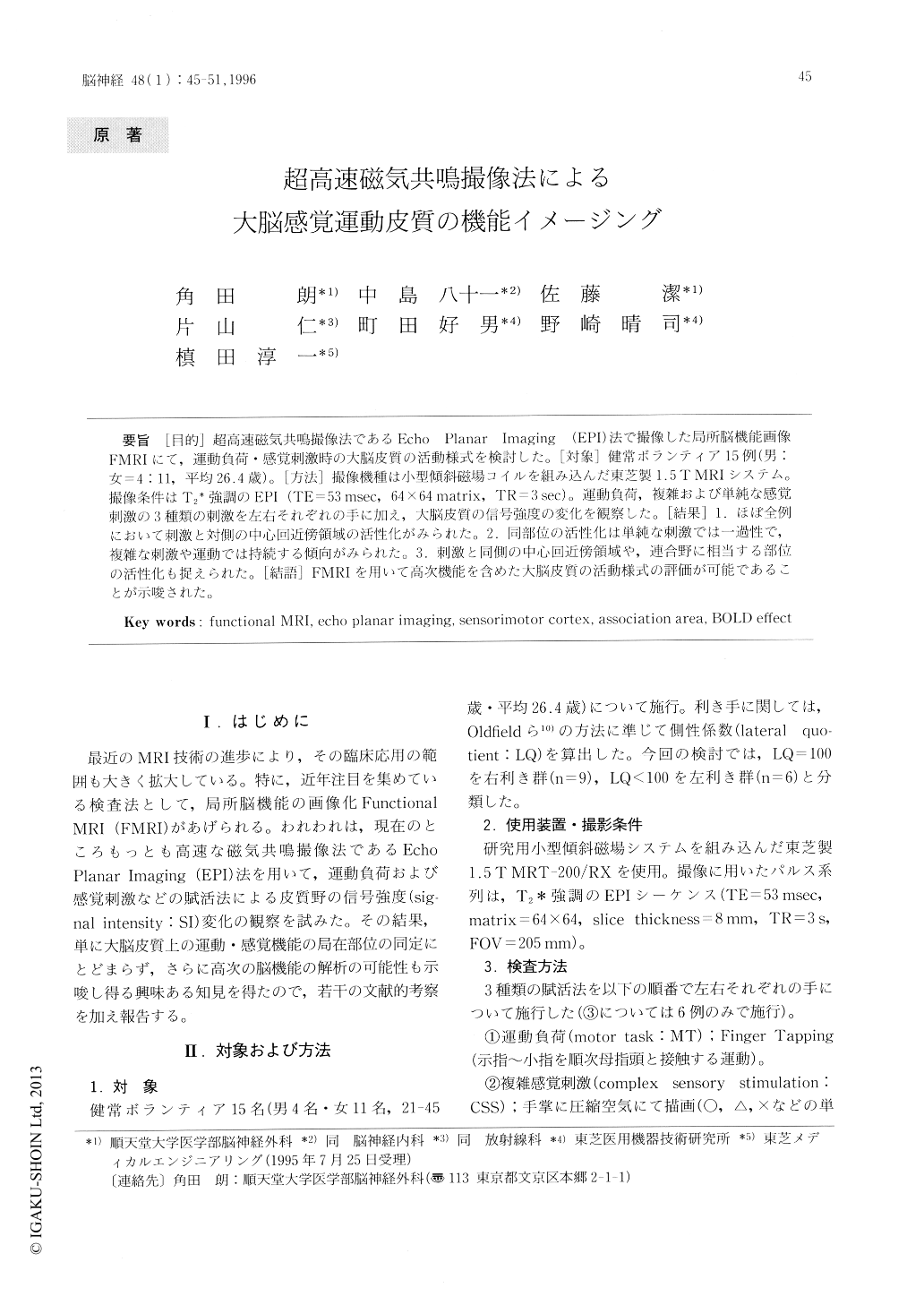
[目的]超高速磁気共鳴撮像法であるEcho Planar Imaging(EPI)法で撮像した局所脳機能画像FMRIにて,運動負荷・感覚刺激時の大脳皮質の活動様式を検討した。[対象]健常ボランティア15例(男:女=4:11,平均26.4歳)。[方法]撮像機種は小型傾斜磁場コイルを組み込んだ東芝製1.5TMRIシステム。撮像条件はT2強調のEPI(TE=53msec,64×64matrix,TR=3sec)。運動負荷,複雑および単純な感覚刺激の3種類の刺激を左右それぞれの手に加え,大脳皮質の信号強度の変化を観察した。[結果]1.ほぼ全例において刺激と対側の中心回近傍領域の活性化がみられた。2.同部位の活性化は単純な刺激では一過性で,複雑な刺激や運動では持続する傾向がみられた。3.刺激と同側の中心回近傍領域や,連合野に相当する部位の活性化も捉えられた。[結語]FMRIを用いて高次機能を含めた大脳皮質の活動様式の野評価が可能であることが示唆された。
The aim of this study was to assess changes in brain activity during a motor task and variable sensory stimulation using echo planar imaging, which represents the fastest clinically usefull imag-ing technique available.
Materials and methods: The subjects of this study were 11 healthy-volunteers, 4 males and 11 felales, with an average of 26.4 years. The subjects were instructed to tap the fingers of one hand as the motor task. Com-pressed air was applied 5 times a second as "simple" sensory stimulation.

Copyright © 1996, Igaku-Shoin Ltd. All rights reserved.


