Japanese
English
- 有料閲覧
- Abstract 文献概要
- 1ページ目 Look Inside
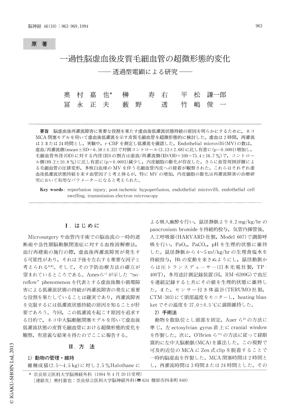
脳虚血後再灌流障害に重要な役割を果たす虚血後低灌流状態持続の原因を明らかにするために,ネコMCA閉塞モデルを用いて虚血後低灌流を示す皮質毛細血管を超微形態的に検討した。虚血は2時間,再灌流は3または24時間とし,実験中,r-CBFを測定し低灌流を確認した。Endothelial microvilli(MV)の数は,虚血/再灌流側(mean±SD=6.58±4.32)で対側コントロール(3.13±2.68)に比し有意に(p=0.0001)増加し,毛細血管外径(OD)に対する内径(ID)の割合は虚血/再灌流側(ID/OD×100=75.4±16.7%)で,コントロール側(89.2±10.8%)に比し有意に(p=0.0001)減少し,内皮細胞の膨化が存在した。さらに血管周囲浮腫による毛細血管の圧排変形,多核白血球のMVを伴う毛細血管内皮への接着が観察された。これらはそれぞれ虚血後低灌流状態持続を来す血管因子と考え得るが,特にMVの増加,内皮細胞の膨化は再灌流障害の治療研究において有用なパラメーターになると考えられた。
Post-ischemic hypoperfusion may play a significant role in reperfusion injury. Since there is no established treatment for hypoperfusion, how-ever we decided to explore the morphological cause of post-ischemic hypoperfusion. In this study we used transmission electron microscopy to investi-gate the capillaries in ischemic/reperfused neocor-tex induced by 2 hours of middle cerebral artery occlusion followed by either 3 or 24 hours of reperfu-sion in 14 cats. Post-ischemic hypoperfusion was confirmed by measuring regional blood flow through a cranial window just above the left ectosylvian gyrus, which has poor anastomosis.

Copyright © 1994, Igaku-Shoin Ltd. All rights reserved.


