Japanese
English
- 有料閲覧
- Abstract 文献概要
- 1ページ目 Look Inside
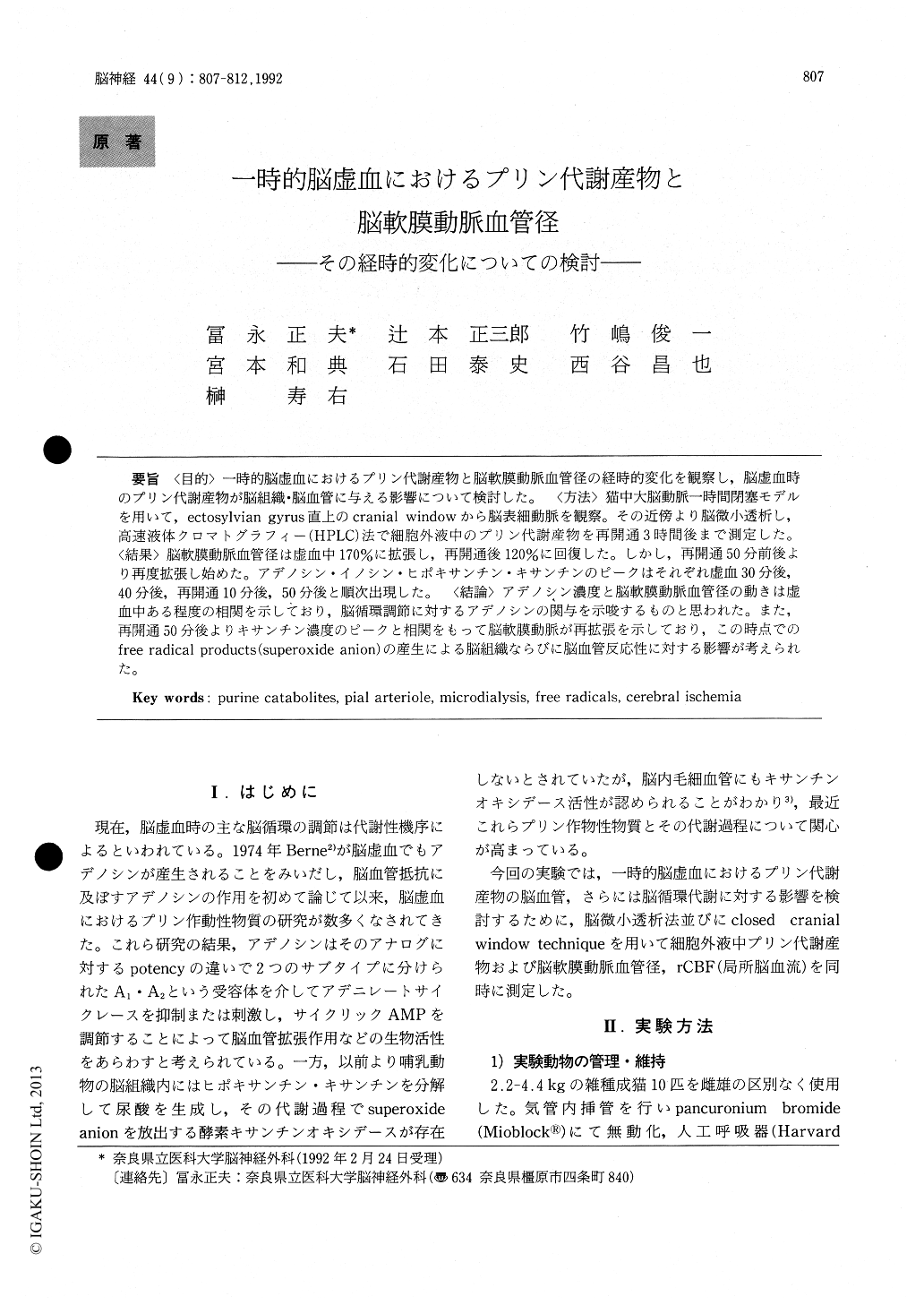
〈目的〉一時的脳虚血におけるプリン代謝産物と脳軟膜動脈血管径の経時的変化を観察し,脳虚血時のプリン代謝産物が脳組織・脳血管に与える影響について検討した。〈方法〉猫中大脳動脈一時間閉塞モデルを用いて,ectosylvian gyrus直上のcranial windowから脳表細動脈を観察。その近傍より脳微小透析し,高速液体クロマトグラフィー(HPLC)法で細胞外液中のプリン代謝産物を再開通3時間後まで測定した。〈結果〉脳軟膜動脈血管径は虚血中170%に拡張し,再開通後120%に回復した。しかし,再開通50分前後より再度拡張し始めた。アデノシン・イノシン・ヒポキサンチン・キサンチンのピークはそれぞれ虚血30分後,40分後,再開通10分後,50分後と順次出現した。〈結論〉アデノシン濃度と脳軟膜動脈血管径の動きは虚血中ある程度の相関を示しており,脳循環調節に対するアデノシンの関与を示唆するものと思われた。また,再開通50分後よりキサンチン濃度のピークと相関をもって脳軟膜動脈が再拡張を示しており,この時点でのfree radical products(superoxide anion)の産生による脳組織ならびに脳血管反応性に対する影響が考えられた。
In transient cerebral ischemia, extracellular pur-ine catabolites and pial arteriolar diameter were measured continuously. Ischemia during one hour was induced by unilateral occlusion of left middle cerebral artery in feline. Extracellular purine catabolites were sampled by in vivo brain mi-crodialysis technique from the gray matter at ectosylvian gyrus. These catabolites were analyzedby HPLC system. Simultaneously, reactivity of pial arteriole was observed and its diameter was mea-sured through the cranial window using intravital microscope and width analyzer.
Extracellular concentrations of adenosine, inosine, hypoxanthine and xanthine were found to be 0.80±0.16μM, 2.01±0.95μM, 4.01±2.73μM and 3.93±2.39μM, respectively. During ischemia, the concentration of adenosine increased 8.7-fold and arteriolar diameter was 170% of the resting state. These findings in extracellular adenosine concentra-tion and pial arteriolar diameter during ischemia support a role of adenosine in regulation of cerebral blood flow.
After reperfusion, arteriolar diameter had return-ed to 120% of the resting state. But 50 min after reperfusion, pial arteriole began to dilate again coincident with the peak of xanthine concentration. These results suggest that free radicals were produced and could affect pial arterioles 50 min after reperfusion.

Copyright © 1992, Igaku-Shoin Ltd. All rights reserved.


