Japanese
English
- 有料閲覧
- Abstract 文献概要
- 1ページ目 Look Inside
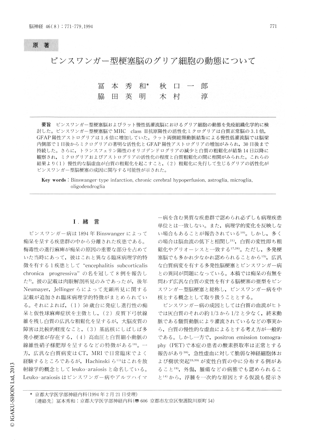
ビンスワンガー型梗塞脳およびラット慢性低灌流脳におけるグリア細胞の動態を免疫組織化学的に検討した。ビンスワンガー型梗塞脳でMHC class II抗原陽性の活性化ミクログリアは白質正常脳の3.1倍,GFAP陽性アストログリアは1.6倍に増加していた。ラット両側総頸動脈結紮による慢性低灌流脳では脳梁内側部で1日後からミクログリアの著明な活性化とGFAP陽性アストログリアの増加がみられ,30日後まで持続した。さらに,トランスフェリン陽性のオリゴデンドログリアの減少と白質の粗鬆化が結紮14日以降に観察され,ミクログリアおよびアストログリアの活性化の程度と白質粗髪化の間に相関がみられた。これらの結果より(1)慢性的な脳虚血が白質の粗鬆化を起こすこと,(2)粗鬆化に先行して生じるグリアの活性化がビンスワンガー型脳梗塞の成因に関与する可能性が示された。
Changes in glial cells were investigated immuno-histochemically in the autopsy brains of patients with Binswanger-type infarction and the brains of rats with chronic cerebral hypoperfusion. Activated microglia, which are positive for MHC class II antigen, and GFAP immunoreactive astroglia were 3.1 times and 1.6 times, respectively, more numer-ous, in Binswanger-type infarction than in normal white matter.

Copyright © 1994, Igaku-Shoin Ltd. All rights reserved.


