Japanese
English
- 有料閲覧
- Abstract 文献概要
- 1ページ目 Look Inside
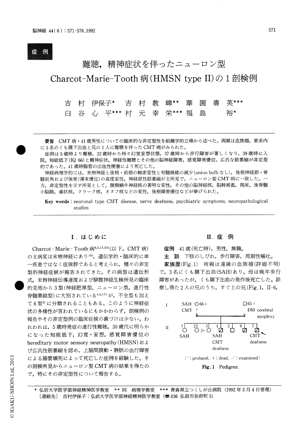
CMT病・41歳男性についての臨床的な非定型性を組織学的立場から述べた。両親は血族婚,家系内に3名のくも膜下出血と兄の1人に難聴を伴ったCMT病がみられた。
症例は5歳時より難聴,32歳時から時々幻覚妄想状態,37歳時から歩行障害が著しくなり,39歳時に入院。知能低下(IQ 66)と精神症状,神経性難聴とその他の脳神経障害,感覚障害優位,広汎な筋萎縮が非定型的であった。41歳時腸管の出血性梗塞により死亡した。
神経病理学的には,末梢神経と後根・前根の軸索変性と有髄線維の減少(onion bulbなし),後根神経節・脊髄前角および後索(薄束優位)の高度変性,神経原性筋萎縮が主所見で,ニューロン型CMT病に一致した。一方,非定型性を示す所見として,腹側蝸牛神経核の著明な変性,その他の脳神経核,脳幹被蓋,視床,後脊髄小脳路,歯状核,クラーク核,オヌフ核などの変性,後根障害優位などが挙げられた。
The clinical and pathological findings of a 41-year-old male patient with atypical Charcot-Marie -Tooth disease were reported. There were 3 cases of subarachnoid haemorrhage, 2 nerve deafness and 2 hereditary motor and sensory neuropathy (HMSN) in his family. He had suffered from prog-ressive nerve deafness since 5 years old and gait disturbance since 37 years old. He had been admit-ted to the psychiatric hospital 3 times because of hallucinatory-delusional state and behavior abnor-malities.
Neurological examinations at 39 years old reve-aled that he had mental deterioration (IQ 66), nerve deafness, diffuse muscle atrophy, most marked dis-tally, sensory disturbance, areflexia, positive Romberg's sign, orthostatic hypotension, dysphagia and slurred speech.
MCV of median nerve was 27.8 m/sec, and SCV was not evoked. EEG revealed nonspecific dysfunc-tion of the brain.
He died of ileus-like condition at 41 years old. General autopsy showed haemorrhagic infarction of the jejunum and ileum due to compression of the superior mesentric artery and vein by an adhesion band of connective tissue formed after previous appendectomy.
Neuropathological examinations revealed axonal degeneration and loss of myelinated fibers with schwannosis of anterior and posterior spinal nerve roots as well as peripheral nerves. The posterior roots were more severely affected than the anterior ones. Ganglion cells of the posterior root ganglia showed remarkable degeneration and loss. There was severe degeneration of the posterior columns, especially in the gracilis, of the spinal cord. Nerve cells in the anterior horns and Clarke's columns also displayed conspicuous atrophy or central chromato-lysis followed by gliosis. There was slight degenera-tion of the posterior spinocerebellar tracts. Central chromatolysis or atrophy of neurons was also noted in the Onuf's nuclei, thalamus and certain nuclei (V, VI, VIII etc) of tegmentum of the brainstem. Of these, choclear nuclei exhibited intense degenera-tion and fibrillary gliosis.
Atypicality as CMT disease was discussed.

Copyright © 1992, Igaku-Shoin Ltd. All rights reserved.


