Japanese
English
- 有料閲覧
- Abstract 文献概要
- 1ページ目 Look Inside
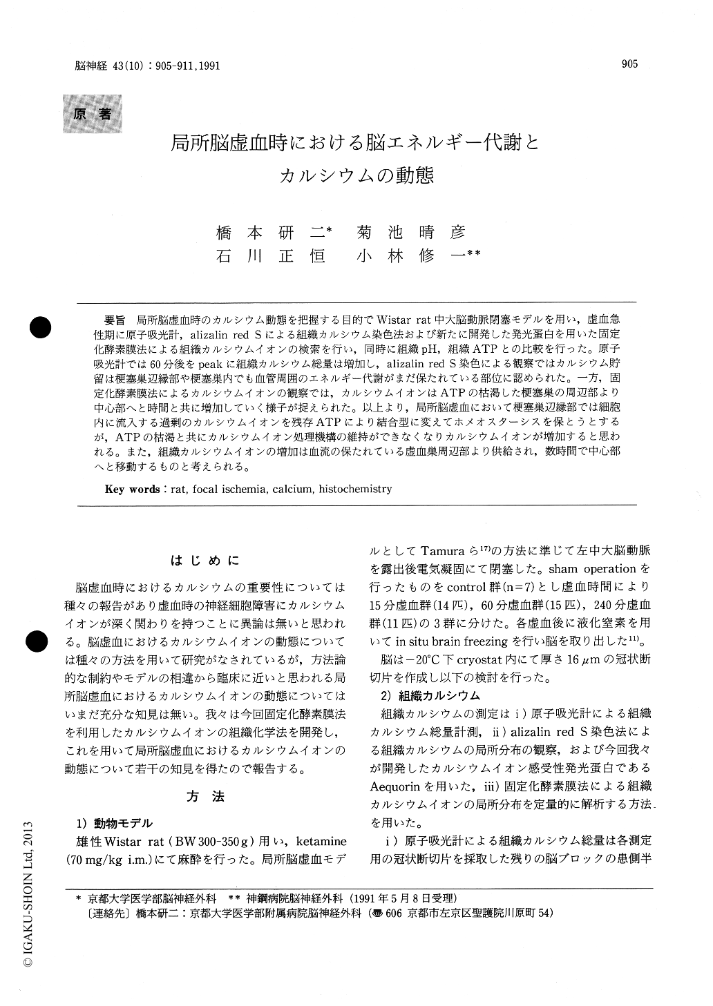
局所脳虚血時のカルシウム動態を把握する目的でWistar rat中大脳動脈閉塞モデルを用い,虚血急性期に原子吸光計,alizalin red Sによる組織カルシウム染色法および新たに開発した発光蛋白を用いた固定化酵素膜法による組織カルシウムイオンの検索を行い,同時に組織pH,組織ATPとの比較を行った。原子吸光計では60分後をpeakに組織カルシウム総量は増加し,alizalin red S染色による観察ではカルシウム貯留は梗塞巣辺縁部や梗塞巣内でも血管周囲のエネルギー代謝がまだ保たれている部位に認められた。一方,固定化酵素膜法によるカルシウムイオンの観察では,カルシウムイオンはATPの枯渇した梗塞巣の周辺部より中心部へと時間と共に増加していく様子が捉えられた。以上より,局所脳虚血において梗塞巣辺縁部では細胞内に流入する過剰のカルシウムイオンを残存ATPにより結合型に変えてホメオスターシスを保とうとするが,ATPの枯渇と共にカルシウムイオン処理機構の維持ができなくなりカルシウムイオンが増加すると思われる。また,組織カルシウムイオンの増加は血流の保たれている虚血巣周辺部より供給され,数時間で中心部へと移動するものと考えられる。
Changes of brain tissue calcium in the focal is-chemia model of Wistar rat were investigated by three different methods ; atomic absorption spectro-photometer, calcium stain with alizarin red S, and new histochemical method using aequorin, a cal-cium ion sensitive photoprotein. Tissue pH and tissue ATP were concomitantly investigated by histochemical method. Rat brain was frozen in situ at 15,60 or 240 minutes after left middle cerebral artery was occluded. Coronal brain sections of 16 μm thickness were made and the brain slices applied for calcium stain and histochemical studies. The residual brain block was applied for atomic absorption spectrophotometric study.
Tissue calcium content of left hemisphere in-creased from 1.34 ± 0.09 (mean±SEM) (n=7) to 1.54±0.16 (n=12), 2.07±0.12 (n=9). 1.69±0.11 (n= 10) μumol/g wet weight after 15,60 and 240 minutes respectively. Calcium stain with alizarin red S showed that the increase of calcium was observed in the peripheral part of the ischemic lesion where ATP was left in a spotty fashion, and calcium deposits disappeared with correspondence to exhaustion of ATP. Tissue calcium ion content studied by newly histochemical method, showed heterogeneous change. At an early stage of theischemia, the increase of tissue calcium ion was shown only in the peripheral part of the ischemic lesion, and it gradually extended to the central part. Calcium ion increased in density in an area corre-sponding to that of the ATP decrease. Within the area of calcium ion increase, regional differences were noted ; a greater increase at the border with the intact area and in the parts where ATP was heterogeneously preserved. In the non-ischemic area close to the ischemic area, where ATP was preserved with mild acidosis, calcium ion decreased more than in the surrounding area where ATP was preserved.
The calcium ion which was influx into the intracellular space by ischemic insult, would be stored in the mitochondria at early stage. A few hours later ATP was exhausted and the tissue calcium ion concentration increased gradually and its increase extended to the central part from the peripheral part, where calcium ions was supplied from the residual blood flow.
In this study, to investigate the regional change of endogeneous culcium ion, we developed a new histo-chemical method using aequorin, a calcium ion sensitive photoprotein. This method is very simple and useful for investigating regional changes of tissue calcium ion on forcal cerebral ischemia.

Copyright © 1991, Igaku-Shoin Ltd. All rights reserved.


