Japanese
English
- 有料閲覧
- Abstract 文献概要
- 1ページ目 Look Inside
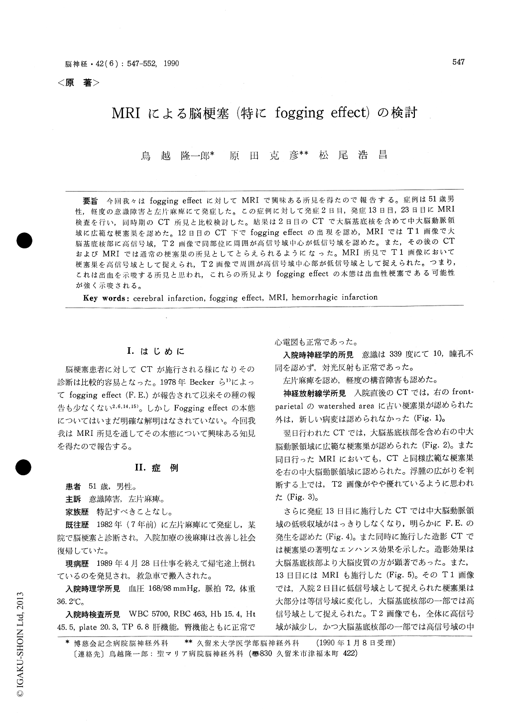
今回我々はfogging effectに対してMRIで興味ある所見を得たので報告する。症例は51歳男性,軽度の意識障害と左片麻痺にて発症した。この症例に対して発症2日目,発症13日目,23日目にMRI検査を行い,同時期のCT所見と比較検討した。結果は2日目のCTで大脳基底核を含めて中大脳動脈領域に広範な梗塞巣を認めた。12日目のCT下でfogging effectの出現を認め,MRIではT1画像で大脳基底核部に高信号域,T2画像で同部位に周囲が高信号域中心が低信号域を認めた。また,その後のCTおよびMRIでは通常の梗塞巣の所見としてとらえられるようになった。MRI所見でT1画像において梗塞巣を高信号域として捉えられ,T2画像で周囲が高信号域巾心部が低信号域として捉えられた。つまり,これは出血を示唆する所見と思われ,これらの所見よりfogging effectの本態は出血性梗塞である可能性が強く示唆される。
Fogging effect in cerebral infarction was studied by MRI in a 51-year-old male patient. Initial symptoms consisted of mild disturbance of con-sciousness and left hemiparesis. MRI examination was performed 2, 13 and 22 days after onset and the results were compared with CT findings during the same period.
CT on day 2 revealed a wide of infarction in the region of the middle cerebral artery including the basal ganglia. The presence of a fogging effect was seen by CT on day 12 and MRI reve-aled a high signal intensity in the region of the basal ganglia in T1 image, a high signal intensity in the peripheral region and a low signal intensity in the center in T2 image. It was possible to define the lesion as the ordinary infarcted lesion by the subsequent CT and MRI. MRI indicated the infarct lesion was to be a high signal intensity in T1 image and a high signal intensity in the periphery and a low signal intensity in the center in T 2 image.
It was concluded that these findings indicated hemorrhage, strongly suggesting that the cause of the fogging effect was hemorrhagic infarction.

Copyright © 1990, Igaku-Shoin Ltd. All rights reserved.


