Japanese
English
- 有料閲覧
- Abstract 文献概要
- 1ページ目 Look Inside
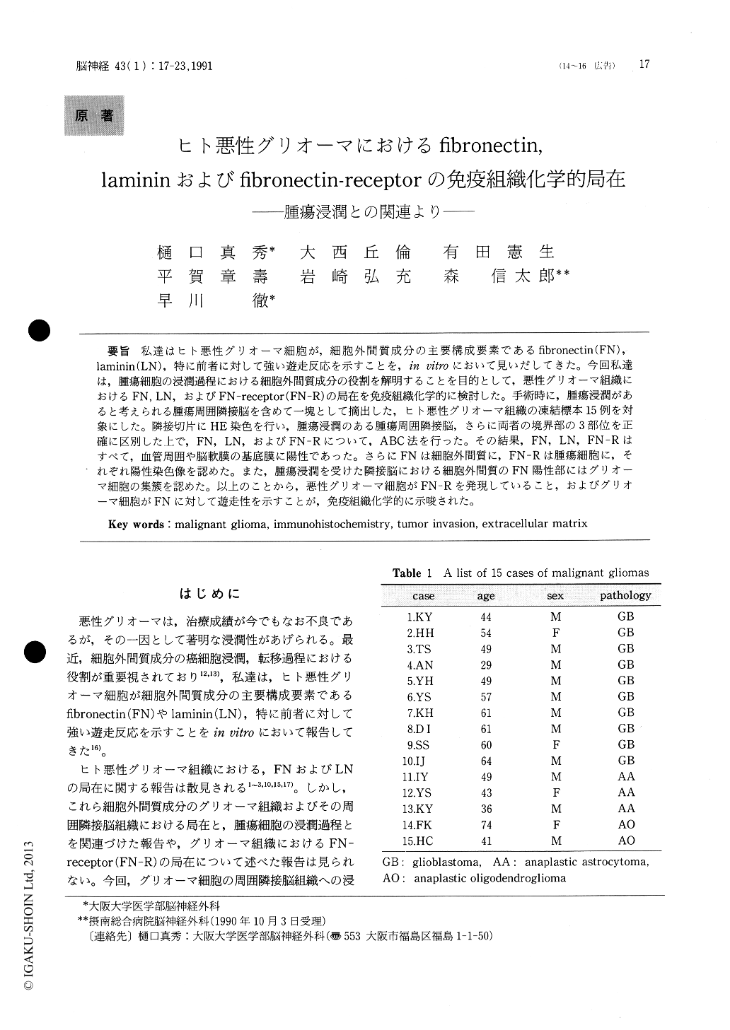
私達はヒト悪性グリオーマ細胞が,細胞外間質成分の主要構成要素であるfibronectin(FN),laminin(LN),特に前者に対して強い遊走反応を示すことを,in vitroにおいて見いだしてきた。今回私達は,腫瘍細胞の浸潤過程における細胞外間質成分の役割を解明することを目的として,悪性グリオーマ組織におけるFN,LN,およびFN-receptor(FN-R)の局在を免疫組織化学的に検討した。手術時に,腫瘍浸潤があると考えられる腫瘍周囲隣接脳を含めて一塊として摘出した,ヒト悪性グリオーマ組織の凍結標本15例を対象にした。隣接切片にHE染色を行い,腫瘍浸潤のある腫瘍周囲隣接脳,さらに両者の境界部の3部位を正確に区別した上で,FN,LN,およびFN-Rについて,ABC法を行った。その結果,FN,LN,FN-Rはすべて,血管周囲や脳軟膜の基底膜に陽性であった。さらにFNは細胞外間質に,FN-Rは腫瘍細胞に,それぞれ陽性染色像を認めた。また,腫瘍浸潤を受けた隣接脳における細胞外間質のFN陽性部にはグリオーマ細胞の集簇を認めた。以上のことから,悪性グリオーマ細胞がFN-Rを発現していること,およびグリオーマ細胞がFNに対して遊走性を示すことが,免疫組織化学的に示唆された。
In order to examine a role of extracellular matrix (ECM) components in the process of glioma cell invasion, we investigated the immunohistochemical localization of fibronectin (FN), laminin (LN) and FN -receptor (FN-R) in human malignant gliomas.
The surgical specimens were obtained from 15 patients with malignant gliomas. Tumor tissue and adjacent brain tissue including tumor infiltration were frozen at -80℃ immediately after the resec-tion. Ten μm thick frozen tissue was cut on a cryostat and divided into three different parts on the histology stained with HE, ie, the tumor region (T), brain tissues with tumor infiltration (I), and the bor-der region between these two parts (B). These sec-tions were air-dried, and fixed with cold acetone (-4℃) for 5min. Adjacent sections were immunohis-tochemically stained by ABC method, using mono-clonal antibody for FN-R and polyclonal antibodies for FN and LN.
FN, LN and FN-R were all stained at the vascu-lar and pial-glial basement membranes intensely in all gliomas. In immunostain for FN, fine networks of FN were observed in the extracellular space in all three parts. Some tumor cells were clustered around such networks of FN in brain tissues with tumor infiltration. Immunostain for LN demonstrat-ed that the vascularity in the border between the tumor and the brain with tumor infiltration was much higher than that in other parts. LN was not stained in the extracellular space in all these gliomas. FN-R was expressed in some tumor cells,especially in the clustered tumor cells in the brain with tumor infiltration.
These findings may indicate that glioma cells can migrate to FN supporting our previous study in vitro of glioma cell migration to FN. In addition, it was suggested that the expression of FN-R in glioma cells might play an important role in the process of glioma cell invasion in the parenchyma of the central nervous system.

Copyright © 1991, Igaku-Shoin Ltd. All rights reserved.


