Japanese
English
- 有料閲覧
- Abstract 文献概要
- 1ページ目 Look Inside
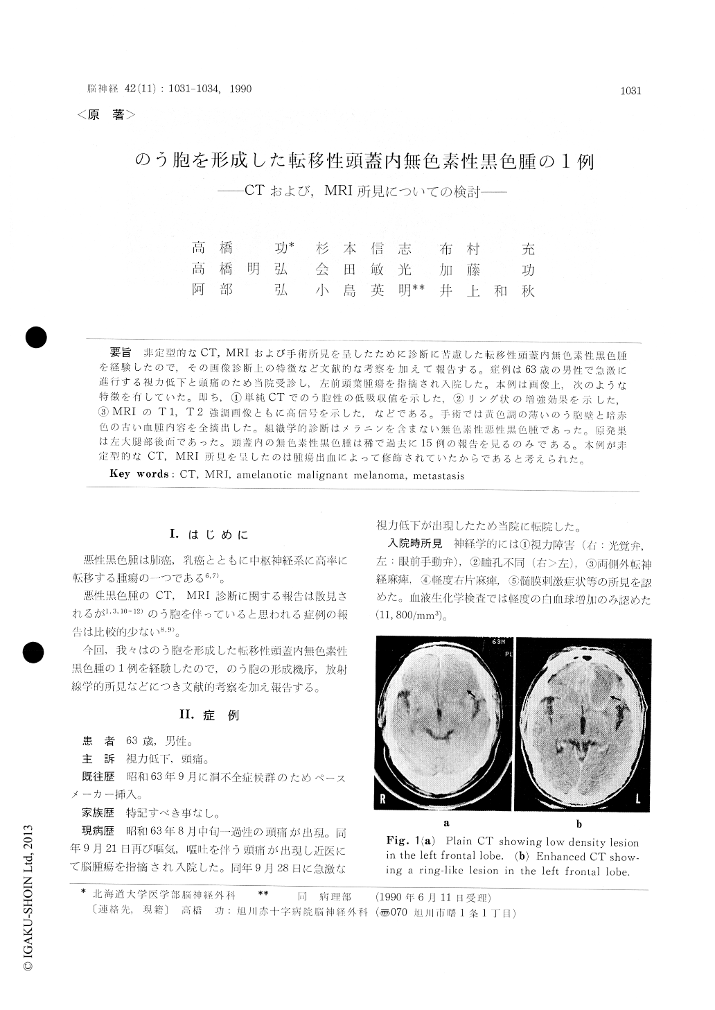
非定型的なCT,MRIおよび手術所見を呈したために診断に苦慮した転移性頭蓋内無色素性黒色腫を経験したので,その画像診断上の特徴など文献的な考察を加えて報告する。症例は63歳の男性で急激に進行する視力低下と頭痛のため当院受診し,左前頭葉腫瘍を指摘され入院した。本例は画像上,次のような特徴を有していた。即ち,①単純CTでのう胞性の低吸収値を示した,②リング状の増強効果を示した,③MRIのT1,T2強調画像ともに高信号を示した,などである。手術では黄色調の薄いのう胞壁と暗赤色の占い血腫内容を全摘出した。組織学的診断はメラニンを含まない無色素性悪性黒色腫であった。原発巣は左大腿部後面であった。頭蓋内の無色素性黒色腫は稀で過去に15例の報告を見るのみである。本例が非定型的なCT,MRI所見を呈したのは腫瘍出血によって修飾されていたからであると考えられた。
A case of cystic intracranial metastatic amelano-tic melanoma is presented. As far as we know, cyst formation in intracranial melanoma is rare, and only 15 cases of intracranial amelanotic mela-noma have been reported until now.
A 63-year-old man was admitted with headache and progressive visual disturbance. CT scan reve-aled a large low-density mass with ring-like enha-ncement in the left frontal lobe. Both T 1-and T 2-weighted MRI images revealed hyperintensity.
A left frontotemporal craniotomy was perform-ed. A yellowish mass was observed in the frontal lobe. The content of the cyst consisted of old he-matoma, xanthochromic fluid and necrotic tissue, was evacuated and the cyst wall was totally resec-ted. No abnormal pigmentation was noted in the cyst wall and surrounding brain tissue.
The histological examination revealed amelano-tic melanoma. Primary lesion was found on the left thigh later and resected. The patient died of further intracranial metastasis with repeated he-morrhage 5 months after the admission.
Both CT and MRI findings of our case is atypical as an intracranial malignant melanoma. However, these are compatible with those of intracerebral hemorrhage in subacute stage. It is suggested that melanoma may make the diagnosis difficult when tumor hemorrhage modifies the images of CT or MRI.

Copyright © 1990, Igaku-Shoin Ltd. All rights reserved.


