Japanese
English
- 有料閲覧
- Abstract 文献概要
- 1ページ目 Look Inside
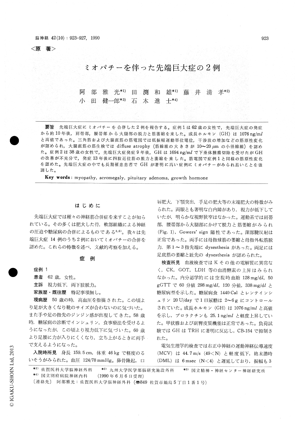
先端巨大症にミオパチーを合併した2例を報告する。症例1は62歳の女性で,先端巨大症の発症から約10年後,肩帯部,腰帯部から大腿部の脱力と筋萎縮を来した。成長ホルモン(GH)は1076 ng/mlと高値であった。三角筋および大腿直筋の筋電図では低振幅運動単位電位,干渉波の増加などの筋原性変化が認められ,大腿直筋の筋生検ではdiffuse atrophy(筋線維の大きさが10〜20μmの小径線維)を認めた。症例2は38歳の女性で,先端巨大症発症9年後,GHは1694 ng/mlで下垂体腫瘍切除を受けたがGHの改善が不充分で,発症13年後に四肢近位筋の脱力と萎縮を来した。筋電図で症例1と同様の筋原性変化を認めた。先端巨大症の中でも長期罹患患者でGHが著明に高い症例にミオパチーがみられ易いことを強調した。
Acromegaly is often associated with neuromus-cular disorders. Most of them are caused by com-pression of nerves with hypertrophic bone and soft tissues or complications of diabetes mellitus. My-opathy has rarely been reported in the Japanese literature. We report two cases with myopathy out of 14 cases of acromegaly.
Case 1 is a 62-year-old woman who developedmuscle weakness and atrophy in the shoulder gir-dle, pelvic girdle and femoral regions after a 10-year history of acromegaly. She showed positive Gowers' sign and normal DTRs. Basal growth hor-mone (GH) level in plasma was 1076 ng/ml. Electro-myograms (EMG) obtained from the deltoid and rectus femoris muscles revealed tyriieal myopathic abnormalities ; an excess of small-amplitude, short-duration, polyphasic motor unit potentials. Histolo-gical examinations of the rectus femoris muscle sho-wed diffuse atrophy of both type I and type II fibers, She also had bilateral carpal tunnel syndrome and bilateral tarsal tunnel syndrome, which were con-firmed by nerve conduction studies of median nerves and posterior tibial nerves. A cranial computed tomography (CT) scan demonstrated sellar mass with suprasellar extension. She underwent transsphenoidal adenomectomy and radiation therapy, GH level lowered to 29 ng/ml, however, myopathy remained unchanged for 3 years after the surgery.
Case 2 is a 38-year-old woman who had undergone partial removal of a pituitary adenoma 9 years after the onset of acromegaly. Basal GH level in plasma before the surgery had been 1694 ng/ml and was still high after the surgery (100-505 ng/ml). The patient developed proximal muscle weakness and atrophy 4years after the surgery. She showed cervico-thora-cic scoliosis. She was normal in DTRs and sensory testing. EMGs revealed myopathic abnormalities.
As neuromuscular complications, we count 2 cases with myopathy and 3 cases with carpal tunel syndro-me out of 14 cases of acromegaly which we recently experienced in our institute. Average durations of acromegaly were 11. 5 years in 2 cases with myopa-thy and 5. 8 years in 12 cases without myopathy. A-verage GH levels were 1385 ng/ml in the former group and 39 ng/ml in the latter group. The diffe-rences were statistically significant (p<0. 05, p< 0.0001). The mechanism of muscle atrophy in GH excess was discussed. A diffuse muscle hypertrophy is usually ihduced in the early stage of GH excess. The hypertrophic muscle, however, is functionally inefficient probably due to reduced membrane excita-bility of muscle fibers or decreased myofibrillar ATP-ase activity. The muscle may be incapable of main-taining the structural integrity of its hypertrophic state in association with disuse of muscles after long duration of GH excess. We emphasize an associa-tion of myopathy in patients with a long duration of acromegaly and very high level of GH.

Copyright © 1990, Igaku-Shoin Ltd. All rights reserved.


